Copper »
PDB 2idq-2pp7 »
2j55 »
Copper in PDB 2j55: X-Ray Reduced Paraccocus Denitrificans Methylamine Dehydrogenase O-Quinone in Complex with Amicyanin.
Enzymatic activity of X-Ray Reduced Paraccocus Denitrificans Methylamine Dehydrogenase O-Quinone in Complex with Amicyanin.
All present enzymatic activity of X-Ray Reduced Paraccocus Denitrificans Methylamine Dehydrogenase O-Quinone in Complex with Amicyanin.:
1.4.99.3;
1.4.99.3;
Protein crystallography data
The structure of X-Ray Reduced Paraccocus Denitrificans Methylamine Dehydrogenase O-Quinone in Complex with Amicyanin., PDB code: 2j55
was solved by
A.R.Pearson,
R.Pahl,
V.L.Davidson,
C.M.Wilmot,
with X-Ray Crystallography technique. A brief refinement statistics is given in the table below:
| Resolution Low / High (Å) | 21.70 / 2.15 |
| Space group | P 41 21 2 |
| Cell size a, b, c (Å), α, β, γ (°) | 122.733, 122.733, 246.591, 90.00, 90.00, 90.00 |
| R / Rfree (%) | 18.9 / 24.5 |
Copper Binding Sites:
The binding sites of Copper atom in the X-Ray Reduced Paraccocus Denitrificans Methylamine Dehydrogenase O-Quinone in Complex with Amicyanin.
(pdb code 2j55). This binding sites where shown within
5.0 Angstroms radius around Copper atom.
In total 2 binding sites of Copper where determined in the X-Ray Reduced Paraccocus Denitrificans Methylamine Dehydrogenase O-Quinone in Complex with Amicyanin., PDB code: 2j55:
Jump to Copper binding site number: 1; 2;
In total 2 binding sites of Copper where determined in the X-Ray Reduced Paraccocus Denitrificans Methylamine Dehydrogenase O-Quinone in Complex with Amicyanin., PDB code: 2j55:
Jump to Copper binding site number: 1; 2;
Copper binding site 1 out of 2 in 2j55
Go back to
Copper binding site 1 out
of 2 in the X-Ray Reduced Paraccocus Denitrificans Methylamine Dehydrogenase O-Quinone in Complex with Amicyanin.
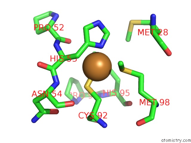
Mono view
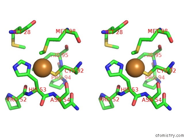
Stereo pair view
Mono view
Stereo pair view
A full contact list of Copper with other atoms in the Cu binding
site number 1 of X-Ray Reduced Paraccocus Denitrificans Methylamine Dehydrogenase O-Quinone in Complex with Amicyanin. within 5.0Å range:
|
Copper binding site 2 out of 2 in 2j55
Go back to
Copper binding site 2 out
of 2 in the X-Ray Reduced Paraccocus Denitrificans Methylamine Dehydrogenase O-Quinone in Complex with Amicyanin.
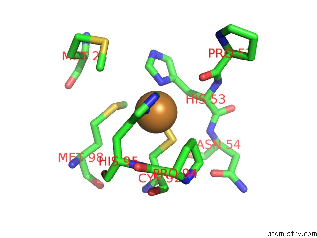
Mono view
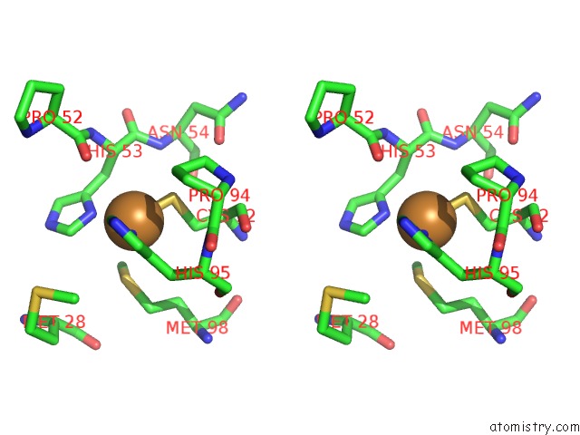
Stereo pair view
Mono view
Stereo pair view
A full contact list of Copper with other atoms in the Cu binding
site number 2 of X-Ray Reduced Paraccocus Denitrificans Methylamine Dehydrogenase O-Quinone in Complex with Amicyanin. within 5.0Å range:
|
Reference:
A.R.Pearson,
R.Pahl,
E.G.Kovaleva,
V.L.Davidson,
C.M.Wilmot.
Tracking X-Ray-Derived Redox Changes in Crystals of A Methylamine Dehydrogenase/Amicyanin Complex Using Single-Crystal Uv/Vis Microspectrophotometry. J.Synchrotron Radiat. V. 14 92 2007.
ISSN: ISSN 0909-0495
PubMed: 17211075
DOI: 10.1107/S0909049506051259
Page generated: Mon Jul 14 01:11:58 2025
ISSN: ISSN 0909-0495
PubMed: 17211075
DOI: 10.1107/S0909049506051259
Last articles
Fe in 2YXOFe in 2YRS
Fe in 2YXC
Fe in 2YNM
Fe in 2YVJ
Fe in 2YP1
Fe in 2YU2
Fe in 2YU1
Fe in 2YQB
Fe in 2YOO