Copper »
PDB 2fqd-2idf »
2i7o »
Copper in PDB 2i7o: Structure of Re(4,7-Dimethyl-Phen)(THR124HIS)(LYS122TRP)(HIS83GLN) Azcu(II), A Rhenium Modified Azurin Mutant
Protein crystallography data
The structure of Structure of Re(4,7-Dimethyl-Phen)(THR124HIS)(LYS122TRP)(HIS83GLN) Azcu(II), A Rhenium Modified Azurin Mutant, PDB code: 2i7o
was solved by
J.Sudhamsu,
B.R.Crane,
with X-Ray Crystallography technique. A brief refinement statistics is given in the table below:
| Resolution Low / High (Å) | 20.00 / 1.50 |
| Space group | I 2 2 2 |
| Cell size a, b, c (Å), α, β, γ (°) | 63.223, 69.075, 68.944, 90.00, 90.00, 90.00 |
| R / Rfree (%) | 23.6 / 25.5 |
Other elements in 2i7o:
The structure of Structure of Re(4,7-Dimethyl-Phen)(THR124HIS)(LYS122TRP)(HIS83GLN) Azcu(II), A Rhenium Modified Azurin Mutant also contains other interesting chemical elements:
| Rhenium | (Re) | 1 atom |
Copper Binding Sites:
The binding sites of Copper atom in the Structure of Re(4,7-Dimethyl-Phen)(THR124HIS)(LYS122TRP)(HIS83GLN) Azcu(II), A Rhenium Modified Azurin Mutant
(pdb code 2i7o). This binding sites where shown within
5.0 Angstroms radius around Copper atom.
In total only one binding site of Copper was determined in the Structure of Re(4,7-Dimethyl-Phen)(THR124HIS)(LYS122TRP)(HIS83GLN) Azcu(II), A Rhenium Modified Azurin Mutant, PDB code: 2i7o:
In total only one binding site of Copper was determined in the Structure of Re(4,7-Dimethyl-Phen)(THR124HIS)(LYS122TRP)(HIS83GLN) Azcu(II), A Rhenium Modified Azurin Mutant, PDB code: 2i7o:
Copper binding site 1 out of 1 in 2i7o
Go back to
Copper binding site 1 out
of 1 in the Structure of Re(4,7-Dimethyl-Phen)(THR124HIS)(LYS122TRP)(HIS83GLN) Azcu(II), A Rhenium Modified Azurin Mutant
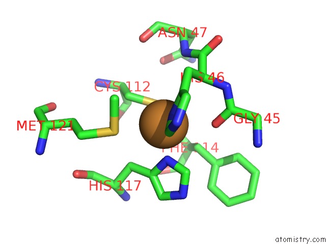
Mono view
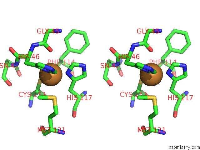
Stereo pair view
Mono view
Stereo pair view
A full contact list of Copper with other atoms in the Cu binding
site number 1 of Structure of Re(4,7-Dimethyl-Phen)(THR124HIS)(LYS122TRP)(HIS83GLN) Azcu(II), A Rhenium Modified Azurin Mutant within 5.0Å range:
|
Reference:
C.Shih,
A.K.Museth,
M.Abrahamsson,
A.M.Blanco-Rodriguez,
A.J.Di Bilio,
J.Sudhamsu,
B.R.Crane,
K.L.Ronayne,
M.Towrie,
A.Vlcek,
J.H.Richards,
J.R.Winkler,
H.B.Gray.
Tryptophan-Accelerated Electron Flow Through Proteins. Science V. 320 1760 2008.
ISSN: ISSN 0036-8075
PubMed: 18583608
DOI: 10.1126/SCIENCE.1158241
Page generated: Mon Jul 14 01:08:36 2025
ISSN: ISSN 0036-8075
PubMed: 18583608
DOI: 10.1126/SCIENCE.1158241
Last articles
F in 4I24F in 4I23
F in 4I0I
F in 4I22
F in 4I0H
F in 4I0J
F in 4HY5
F in 4HY6
F in 4HXN
F in 4HT2