Copper »
PDB 2fqd-2idf »
2hh7 »
Copper in PDB 2hh7: Crystal Structure of Cu(I) Bound Csor From Mycobacterium Tuberculosis.
Protein crystallography data
The structure of Crystal Structure of Cu(I) Bound Csor From Mycobacterium Tuberculosis., PDB code: 2hh7
was solved by
J.C.Sacchettini,
A.Ramesh,
with X-Ray Crystallography technique. A brief refinement statistics is given in the table below:
| Resolution Low / High (Å) | 20.00 / 2.55 |
| Space group | P 64 2 2 |
| Cell size a, b, c (Å), α, β, γ (°) | 91.059, 91.059, 46.779, 90.00, 90.00, 120.00 |
| R / Rfree (%) | 23.1 / 27.9 |
Copper Binding Sites:
The binding sites of Copper atom in the Crystal Structure of Cu(I) Bound Csor From Mycobacterium Tuberculosis.
(pdb code 2hh7). This binding sites where shown within
5.0 Angstroms radius around Copper atom.
In total only one binding site of Copper was determined in the Crystal Structure of Cu(I) Bound Csor From Mycobacterium Tuberculosis., PDB code: 2hh7:
In total only one binding site of Copper was determined in the Crystal Structure of Cu(I) Bound Csor From Mycobacterium Tuberculosis., PDB code: 2hh7:
Copper binding site 1 out of 1 in 2hh7
Go back to
Copper binding site 1 out
of 1 in the Crystal Structure of Cu(I) Bound Csor From Mycobacterium Tuberculosis.
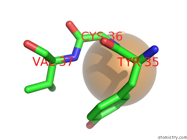
Mono view
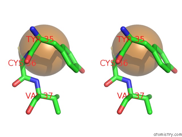
Stereo pair view
Mono view
Stereo pair view
A full contact list of Copper with other atoms in the Cu binding
site number 1 of Crystal Structure of Cu(I) Bound Csor From Mycobacterium Tuberculosis. within 5.0Å range:
|
Reference:
T.Liu,
A.Ramesh,
Z.Ma,
S.K.Ward,
L.Zhang,
G.N.George,
A.M.Talaat,
J.C.Sacchettini,
D.P.Giedroc.
Csor Is A Novel Mycobacterium Tuberculosis Copper-Sensing Transcriptional Regulator. Nat.Chem.Biol. V. 3 60 2007.
ISSN: ISSN 1552-4450
PubMed: 17143269
DOI: 10.1038/NCHEMBIO844
Page generated: Mon Jul 14 01:06:45 2025
ISSN: ISSN 1552-4450
PubMed: 17143269
DOI: 10.1038/NCHEMBIO844
Last articles
Fe in 2YXOFe in 2YRS
Fe in 2YXC
Fe in 2YNM
Fe in 2YVJ
Fe in 2YP1
Fe in 2YU2
Fe in 2YU1
Fe in 2YQB
Fe in 2YOO