Copper »
PDB 2fqd-2idf »
2gi0 »
Copper in PDB 2gi0: Crystal Structure of Cu(I) PHE114PRO Azurin Mutant
Protein crystallography data
The structure of Crystal Structure of Cu(I) PHE114PRO Azurin Mutant, PDB code: 2gi0
was solved by
S.Yanagisawa,
M.J.Banfield,
C.Dennison,
with X-Ray Crystallography technique. A brief refinement statistics is given in the table below:
| Resolution Low / High (Å) | 47.80 / 1.70 |
| Space group | P 21 21 21 |
| Cell size a, b, c (Å), α, β, γ (°) | 35.390, 47.860, 132.550, 90.00, 90.00, 90.00 |
| R / Rfree (%) | 17.9 / 22.4 |
Copper Binding Sites:
The binding sites of Copper atom in the Crystal Structure of Cu(I) PHE114PRO Azurin Mutant
(pdb code 2gi0). This binding sites where shown within
5.0 Angstroms radius around Copper atom.
In total 2 binding sites of Copper where determined in the Crystal Structure of Cu(I) PHE114PRO Azurin Mutant, PDB code: 2gi0:
Jump to Copper binding site number: 1; 2;
In total 2 binding sites of Copper where determined in the Crystal Structure of Cu(I) PHE114PRO Azurin Mutant, PDB code: 2gi0:
Jump to Copper binding site number: 1; 2;
Copper binding site 1 out of 2 in 2gi0
Go back to
Copper binding site 1 out
of 2 in the Crystal Structure of Cu(I) PHE114PRO Azurin Mutant
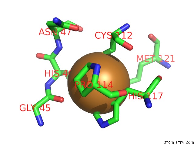
Mono view
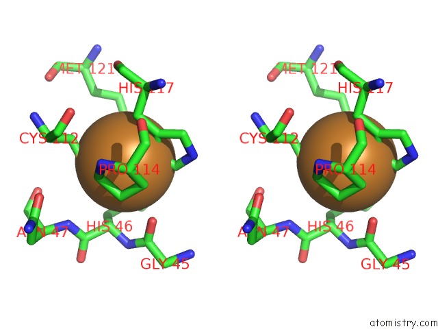
Stereo pair view
Mono view
Stereo pair view
A full contact list of Copper with other atoms in the Cu binding
site number 1 of Crystal Structure of Cu(I) PHE114PRO Azurin Mutant within 5.0Å range:
|
Copper binding site 2 out of 2 in 2gi0
Go back to
Copper binding site 2 out
of 2 in the Crystal Structure of Cu(I) PHE114PRO Azurin Mutant
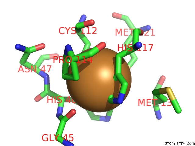
Mono view
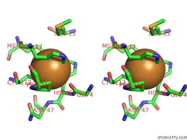
Stereo pair view
Mono view
Stereo pair view
A full contact list of Copper with other atoms in the Cu binding
site number 2 of Crystal Structure of Cu(I) PHE114PRO Azurin Mutant within 5.0Å range:
|
Reference:
S.Yanagisawa,
M.J.Banfield,
C.Dennison.
The Role of Hydrogen Bonding at the Active Site of A Cupredoxin: the PHE114PRO Azurin Variant. Biochemistry V. 45 8812 2006.
ISSN: ISSN 0006-2960
PubMed: 16846224
DOI: 10.1021/BI0606851
Page generated: Mon Jul 14 01:05:23 2025
ISSN: ISSN 0006-2960
PubMed: 16846224
DOI: 10.1021/BI0606851
Last articles
Fe in 2YXOFe in 2YRS
Fe in 2YXC
Fe in 2YNM
Fe in 2YVJ
Fe in 2YP1
Fe in 2YU2
Fe in 2YU1
Fe in 2YQB
Fe in 2YOO