Copper »
PDB 2fqd-2idf »
2gba »
Copper in PDB 2gba: Reduced Cu(I) Form at pH 4 of P52G Mutant of Amicyanin
Protein crystallography data
The structure of Reduced Cu(I) Form at pH 4 of P52G Mutant of Amicyanin, PDB code: 2gba
was solved by
J.K.Ma,
C.J.Carrell,
F.S.Mathews,
V.L.Davidson,
with X-Ray Crystallography technique. A brief refinement statistics is given in the table below:
| Resolution Low / High (Å) | 20.00 / 0.92 |
| Space group | P 1 21 1 |
| Cell size a, b, c (Å), α, β, γ (°) | 28.450, 56.132, 27.003, 90.00, 96.98, 90.00 |
| R / Rfree (%) | 10.8 / 14.8 |
Copper Binding Sites:
The binding sites of Copper atom in the Reduced Cu(I) Form at pH 4 of P52G Mutant of Amicyanin
(pdb code 2gba). This binding sites where shown within
5.0 Angstroms radius around Copper atom.
In total 2 binding sites of Copper where determined in the Reduced Cu(I) Form at pH 4 of P52G Mutant of Amicyanin, PDB code: 2gba:
Jump to Copper binding site number: 1; 2;
In total 2 binding sites of Copper where determined in the Reduced Cu(I) Form at pH 4 of P52G Mutant of Amicyanin, PDB code: 2gba:
Jump to Copper binding site number: 1; 2;
Copper binding site 1 out of 2 in 2gba
Go back to
Copper binding site 1 out
of 2 in the Reduced Cu(I) Form at pH 4 of P52G Mutant of Amicyanin
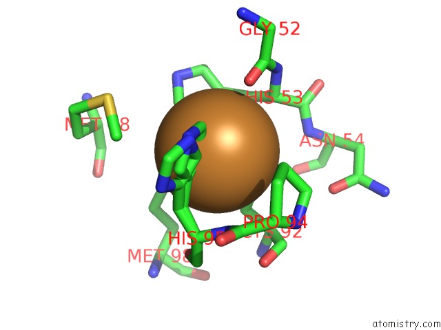
Mono view
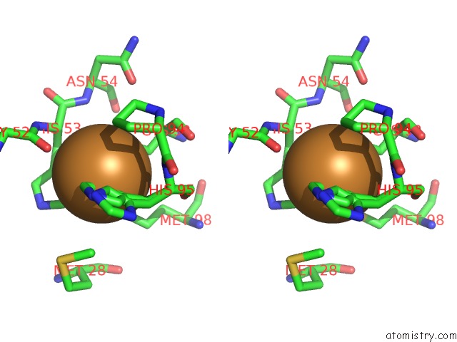
Stereo pair view
Mono view
Stereo pair view
A full contact list of Copper with other atoms in the Cu binding
site number 1 of Reduced Cu(I) Form at pH 4 of P52G Mutant of Amicyanin within 5.0Å range:
|
Copper binding site 2 out of 2 in 2gba
Go back to
Copper binding site 2 out
of 2 in the Reduced Cu(I) Form at pH 4 of P52G Mutant of Amicyanin
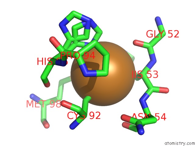
Mono view
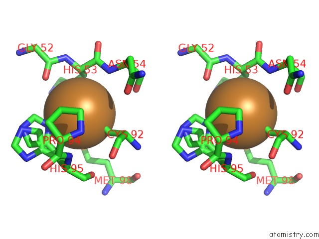
Stereo pair view
Mono view
Stereo pair view
A full contact list of Copper with other atoms in the Cu binding
site number 2 of Reduced Cu(I) Form at pH 4 of P52G Mutant of Amicyanin within 5.0Å range:
|
Reference:
J.K.Ma,
C.J.Carrell,
F.S.Mathews,
V.L.Davidson.
Site-Directed Mutagenesis of Proline 52 to Glycine in Amicyanin Converts A True Electron Transfer Reaction Into One That Is Conformationally Gated. Biochemistry V. 45 8284 2006.
ISSN: ISSN 0006-2960
PubMed: 16819827
DOI: 10.1021/BI0605134
Page generated: Mon Jul 14 01:04:23 2025
ISSN: ISSN 0006-2960
PubMed: 16819827
DOI: 10.1021/BI0605134
Last articles
F in 4DBUF in 4DHM
F in 4DEB
F in 4DC3
F in 4D8C
F in 4D83
F in 4DBQ
F in 4DBN
F in 4DAN
F in 4DA4