Copper in PDB 2ein: Zinc Ion Binding Structure of Bovine Heart Cytochrome C Oxidase in the Fully Oxidized State
Enzymatic activity of Zinc Ion Binding Structure of Bovine Heart Cytochrome C Oxidase in the Fully Oxidized State
All present enzymatic activity of Zinc Ion Binding Structure of Bovine Heart Cytochrome C Oxidase in the Fully Oxidized State:
1.9.3.1;
Protein crystallography data
The structure of Zinc Ion Binding Structure of Bovine Heart Cytochrome C Oxidase in the Fully Oxidized State, PDB code: 2ein
was solved by
K.Muramoto,
K.Hirata,
K.Shinzawa-Itoh,
S.Yoko-O,
E.Yamashita,
H.Aoyama,
T.Tsukihara,
S.Yoshikawa,
with X-Ray Crystallography technique. A brief refinement statistics is given in the table below:
Resolution Low / High (Å)
|
40.00 /
2.70
|
Space group
|
P 21 21 21
|
Cell size a, b, c (Å), α, β, γ (°)
|
187.810,
203.581,
177.927,
90.00,
90.00,
90.00
|
R / Rfree (%)
|
20.8 /
25.8
|
Other elements in 2ein:
The structure of Zinc Ion Binding Structure of Bovine Heart Cytochrome C Oxidase in the Fully Oxidized State also contains other interesting chemical elements:
Copper Binding Sites:
The binding sites of Copper atom in the Zinc Ion Binding Structure of Bovine Heart Cytochrome C Oxidase in the Fully Oxidized State
(pdb code 2ein). This binding sites where shown within
5.0 Angstroms radius around Copper atom.
In total 6 binding sites of Copper where determined in the
Zinc Ion Binding Structure of Bovine Heart Cytochrome C Oxidase in the Fully Oxidized State, PDB code: 2ein:
Jump to Copper binding site number:
1;
2;
3;
4;
5;
6;
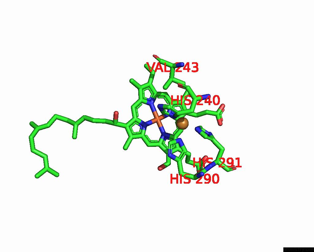
Copper binding site 1 out
of 6 in 2ein
Go back to
Copper Binding Sites List in 2ein
Copper binding site 1 out
of 6 in the Zinc Ion Binding Structure of Bovine Heart Cytochrome C Oxidase in the Fully Oxidized State
Mono view
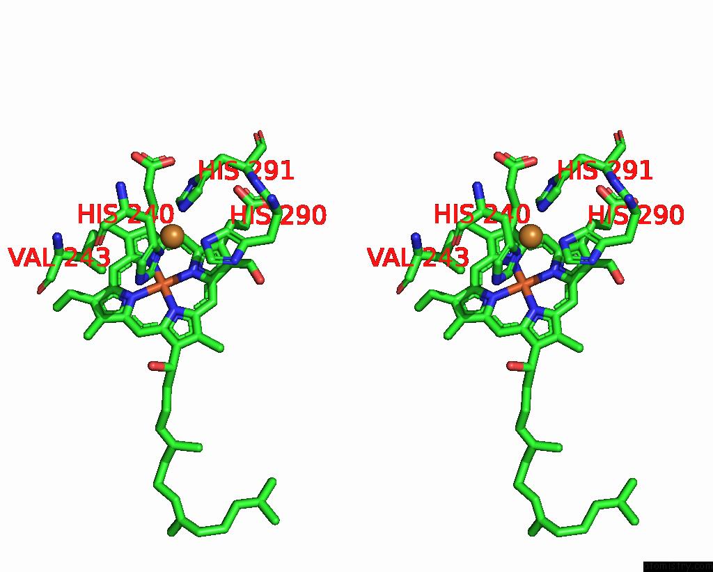
Stereo pair view
|
A full contact list of Copper with other atoms in the Cu binding
site number 1 of Zinc Ion Binding Structure of Bovine Heart Cytochrome C Oxidase in the Fully Oxidized State within 5.0Å range:
probe
|
atom
|
residue
|
distance (Å)
|
B
|
Occ
|
A:Cu517
b:41.4
occ:1.00
|
ND1
|
A:HIS240
|
1.9
|
48.0
|
1.0
|
NE2
|
A:HIS291
|
2.0
|
57.0
|
1.0
|
NE2
|
A:HIS290
|
2.0
|
45.5
|
1.0
|
CE1
|
A:HIS240
|
2.8
|
45.9
|
1.0
|
CE1
|
A:HIS290
|
2.9
|
49.1
|
1.0
|
CE1
|
A:HIS291
|
3.0
|
60.3
|
1.0
|
CD2
|
A:HIS291
|
3.0
|
55.8
|
1.0
|
CG
|
A:HIS240
|
3.1
|
39.0
|
1.0
|
CD2
|
A:HIS290
|
3.2
|
46.1
|
1.0
|
CB
|
A:HIS240
|
3.5
|
34.0
|
1.0
|
CA
|
A:HIS240
|
3.8
|
31.6
|
1.0
|
NE2
|
A:HIS240
|
4.0
|
43.6
|
1.0
|
ND1
|
A:HIS290
|
4.1
|
47.8
|
1.0
|
CD2
|
A:HIS240
|
4.1
|
41.6
|
1.0
|
ND1
|
A:HIS291
|
4.1
|
51.3
|
1.0
|
CG
|
A:HIS291
|
4.2
|
47.9
|
1.0
|
CG
|
A:HIS290
|
4.2
|
47.0
|
1.0
|
NA
|
A:HEA516
|
4.4
|
50.4
|
1.0
|
C1A
|
A:HEA516
|
4.4
|
45.4
|
1.0
|
CG2
|
A:VAL243
|
4.6
|
31.4
|
1.0
|
FE
|
A:HEA516
|
4.6
|
34.0
|
1.0
|
C2A
|
A:HEA516
|
4.7
|
41.3
|
1.0
|
C4A
|
A:HEA516
|
4.8
|
48.6
|
1.0
|
N
|
A:HIS240
|
4.8
|
29.4
|
1.0
|
CHA
|
A:HEA516
|
4.8
|
45.6
|
1.0
|
O
|
A:HIS240
|
4.8
|
36.8
|
1.0
|
C3A
|
A:HEA516
|
4.9
|
43.7
|
1.0
|
C
|
A:HIS240
|
4.9
|
36.2
|
1.0
|
CG1
|
A:VAL243
|
4.9
|
36.4
|
1.0
|
ND
|
A:HEA516
|
4.9
|
41.8
|
1.0
|
|
Copper binding site 2 out
of 6 in 2ein
Go back to
Copper Binding Sites List in 2ein
Copper binding site 2 out
of 6 in the Zinc Ion Binding Structure of Bovine Heart Cytochrome C Oxidase in the Fully Oxidized State
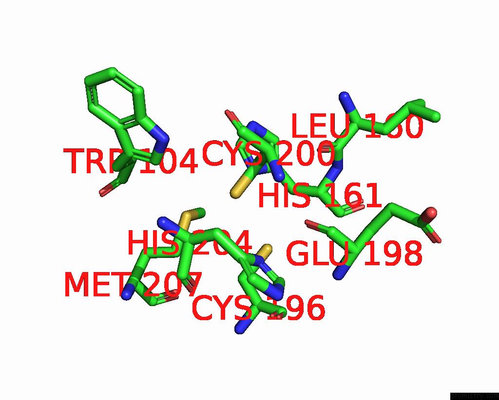
Mono view
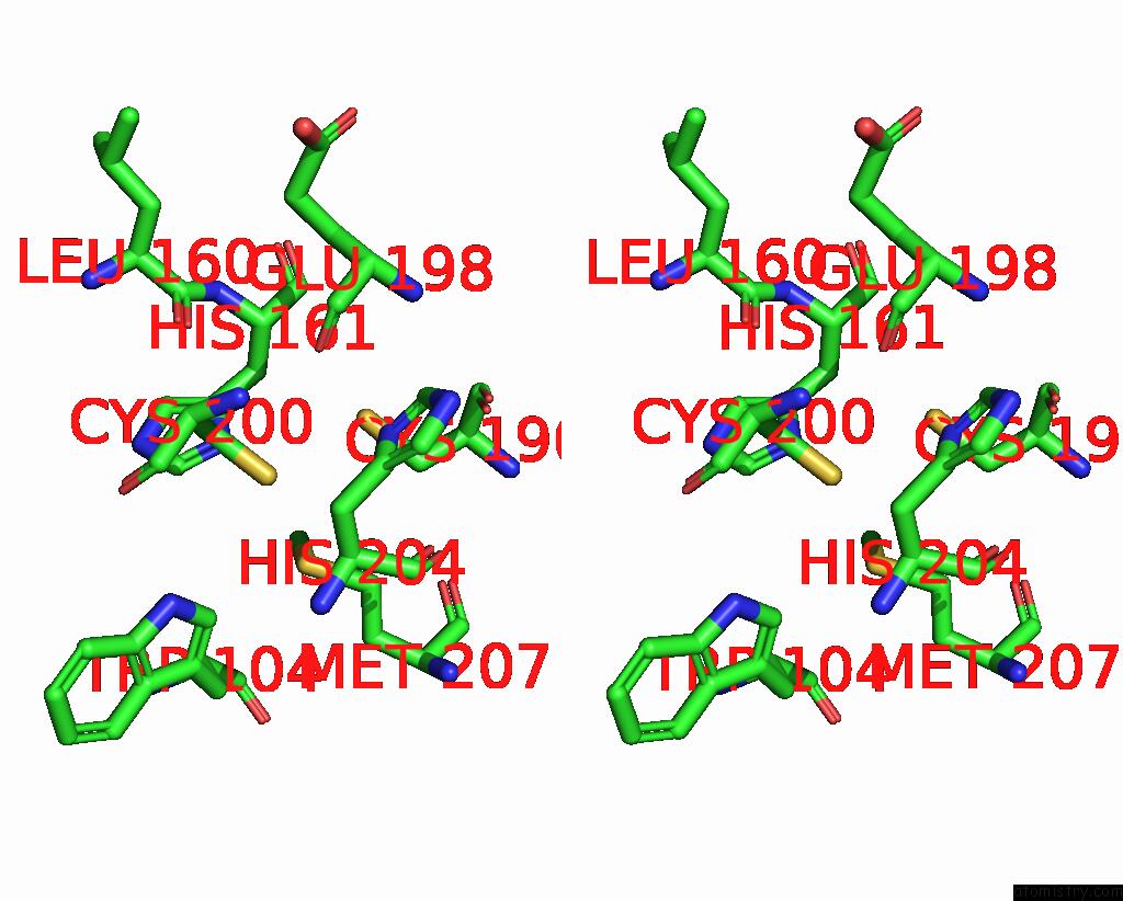
Stereo pair view
|
A full contact list of Copper with other atoms in the Cu binding
site number 2 of Zinc Ion Binding Structure of Bovine Heart Cytochrome C Oxidase in the Fully Oxidized State within 5.0Å range:
probe
|
atom
|
residue
|
distance (Å)
|
B
|
Occ
|
B:Cu228
b:44.6
occ:1.00
|
CU1
|
B:CUA228
|
0.0
|
44.6
|
1.0
|
ND1
|
B:HIS161
|
2.1
|
25.7
|
1.0
|
SG
|
B:CYS196
|
2.3
|
32.5
|
1.0
|
SG
|
B:CYS200
|
2.4
|
27.7
|
1.0
|
CU2
|
B:CUA228
|
2.5
|
41.9
|
1.0
|
SD
|
B:MET207
|
2.8
|
44.8
|
1.0
|
CE1
|
B:HIS161
|
3.1
|
21.8
|
1.0
|
CB
|
B:CYS200
|
3.1
|
36.8
|
1.0
|
CG
|
B:HIS161
|
3.1
|
19.7
|
1.0
|
CE
|
B:MET207
|
3.2
|
32.0
|
1.0
|
CB
|
B:CYS196
|
3.4
|
31.7
|
1.0
|
CB
|
B:HIS161
|
3.4
|
24.3
|
1.0
|
CG
|
B:MET207
|
3.9
|
37.7
|
1.0
|
O
|
B:GLU198
|
4.0
|
49.4
|
1.0
|
CA
|
B:HIS161
|
4.0
|
27.0
|
1.0
|
CD2
|
B:HIS161
|
4.2
|
24.4
|
1.0
|
NE2
|
B:HIS161
|
4.2
|
23.6
|
1.0
|
ND1
|
B:HIS204
|
4.5
|
48.1
|
1.0
|
CA
|
B:CYS200
|
4.5
|
43.1
|
1.0
|
O
|
B:LEU160
|
4.6
|
39.5
|
1.0
|
CA
|
B:CYS196
|
4.7
|
38.2
|
1.0
|
N
|
B:CYS200
|
4.8
|
45.8
|
1.0
|
CA
|
B:HIS204
|
4.9
|
44.1
|
1.0
|
CD1
|
B:TRP104
|
5.0
|
47.1
|
1.0
|
|
Copper binding site 3 out
of 6 in 2ein
Go back to
Copper Binding Sites List in 2ein
Copper binding site 3 out
of 6 in the Zinc Ion Binding Structure of Bovine Heart Cytochrome C Oxidase in the Fully Oxidized State
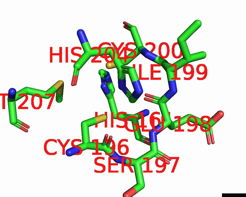
Mono view
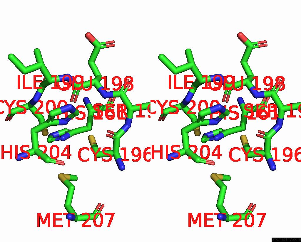
Stereo pair view
|
A full contact list of Copper with other atoms in the Cu binding
site number 3 of Zinc Ion Binding Structure of Bovine Heart Cytochrome C Oxidase in the Fully Oxidized State within 5.0Å range:
probe
|
atom
|
residue
|
distance (Å)
|
B
|
Occ
|
B:Cu228
b:41.9
occ:1.00
|
CU2
|
B:CUA228
|
0.0
|
41.9
|
1.0
|
ND1
|
B:HIS204
|
2.0
|
48.1
|
1.0
|
SG
|
B:CYS200
|
2.2
|
27.7
|
1.0
|
SG
|
B:CYS196
|
2.3
|
32.5
|
1.0
|
O
|
B:GLU198
|
2.4
|
49.4
|
1.0
|
CU1
|
B:CUA228
|
2.5
|
44.6
|
1.0
|
CE1
|
B:HIS204
|
2.9
|
44.4
|
1.0
|
CG
|
B:HIS204
|
3.0
|
45.2
|
1.0
|
CB
|
B:CYS196
|
3.1
|
31.7
|
1.0
|
CB
|
B:CYS200
|
3.4
|
36.8
|
1.0
|
CB
|
B:HIS204
|
3.4
|
42.6
|
1.0
|
C
|
B:GLU198
|
3.5
|
39.2
|
1.0
|
CA
|
B:HIS204
|
3.6
|
44.1
|
1.0
|
N
|
B:CYS200
|
3.6
|
45.8
|
1.0
|
O
|
B:HIS204
|
3.8
|
40.3
|
1.0
|
O
|
B:CYS196
|
3.8
|
48.4
|
1.0
|
N
|
B:GLU198
|
4.0
|
31.4
|
1.0
|
C
|
B:HIS204
|
4.1
|
42.1
|
1.0
|
C
|
B:CYS196
|
4.1
|
45.2
|
1.0
|
NE2
|
B:HIS204
|
4.1
|
41.8
|
1.0
|
C
|
B:ILE199
|
4.1
|
45.8
|
1.0
|
CA
|
B:CYS200
|
4.2
|
43.1
|
1.0
|
CD2
|
B:HIS204
|
4.2
|
42.4
|
1.0
|
N
|
B:ILE199
|
4.2
|
40.6
|
1.0
|
CA
|
B:ILE199
|
4.2
|
43.8
|
1.0
|
CA
|
B:CYS196
|
4.3
|
38.2
|
1.0
|
ND1
|
B:HIS161
|
4.3
|
25.7
|
1.0
|
CA
|
B:GLU198
|
4.4
|
33.5
|
1.0
|
SD
|
B:MET207
|
4.4
|
44.8
|
1.0
|
N
|
B:SER197
|
4.8
|
44.0
|
1.0
|
CG
|
B:MET207
|
4.8
|
37.7
|
1.0
|
N
|
B:HIS204
|
4.9
|
43.0
|
1.0
|
O
|
B:ILE199
|
4.9
|
42.1
|
1.0
|
CA
|
B:HIS161
|
5.0
|
27.0
|
1.0
|
|
Copper binding site 4 out
of 6 in 2ein
Go back to
Copper Binding Sites List in 2ein
Copper binding site 4 out
of 6 in the Zinc Ion Binding Structure of Bovine Heart Cytochrome C Oxidase in the Fully Oxidized State
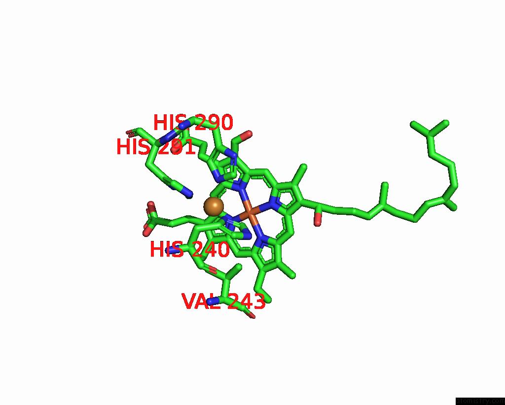
Mono view
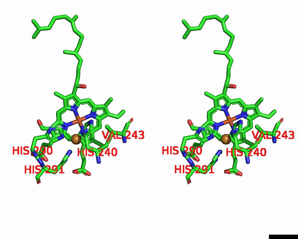
Stereo pair view
|
A full contact list of Copper with other atoms in the Cu binding
site number 4 of Zinc Ion Binding Structure of Bovine Heart Cytochrome C Oxidase in the Fully Oxidized State within 5.0Å range:
probe
|
atom
|
residue
|
distance (Å)
|
B
|
Occ
|
N:Cu517
b:48.3
occ:1.00
|
NE2
|
N:HIS291
|
1.9
|
44.0
|
1.0
|
ND1
|
N:HIS240
|
2.0
|
48.4
|
1.0
|
NE2
|
N:HIS290
|
2.0
|
39.5
|
1.0
|
CE1
|
N:HIS291
|
2.4
|
46.4
|
1.0
|
CE1
|
N:HIS240
|
2.8
|
46.6
|
1.0
|
CE1
|
N:HIS290
|
3.0
|
48.4
|
1.0
|
CG
|
N:HIS240
|
3.1
|
41.3
|
1.0
|
CD2
|
N:HIS290
|
3.2
|
43.4
|
1.0
|
CD2
|
N:HIS291
|
3.3
|
52.2
|
1.0
|
CB
|
N:HIS240
|
3.6
|
37.8
|
1.0
|
ND1
|
N:HIS291
|
3.7
|
36.0
|
1.0
|
NE2
|
N:HIS240
|
4.0
|
42.2
|
1.0
|
CA
|
N:HIS240
|
4.0
|
37.3
|
1.0
|
CG
|
N:HIS291
|
4.1
|
47.1
|
1.0
|
CD2
|
N:HIS240
|
4.1
|
42.3
|
1.0
|
ND1
|
N:HIS290
|
4.2
|
39.7
|
1.0
|
NA
|
N:HEA516
|
4.2
|
49.4
|
1.0
|
CG
|
N:HIS290
|
4.3
|
42.4
|
1.0
|
C1A
|
N:HEA516
|
4.3
|
49.3
|
1.0
|
FE
|
N:HEA516
|
4.3
|
43.6
|
1.0
|
CG2
|
N:VAL243
|
4.6
|
49.0
|
1.0
|
C2A
|
N:HEA516
|
4.6
|
50.3
|
1.0
|
C4A
|
N:HEA516
|
4.7
|
50.1
|
1.0
|
ND
|
N:HEA516
|
4.7
|
43.7
|
1.0
|
CG1
|
N:VAL243
|
4.7
|
56.0
|
1.0
|
CHA
|
N:HEA516
|
4.7
|
42.3
|
1.0
|
C3A
|
N:HEA516
|
4.8
|
49.8
|
1.0
|
NB
|
N:HEA516
|
4.9
|
44.1
|
1.0
|
N
|
N:HIS240
|
4.9
|
33.6
|
1.0
|
C4D
|
N:HEA516
|
4.9
|
42.7
|
1.0
|
O
|
N:HIS240
|
5.0
|
46.4
|
1.0
|
|
Copper binding site 5 out
of 6 in 2ein
Go back to
Copper Binding Sites List in 2ein
Copper binding site 5 out
of 6 in the Zinc Ion Binding Structure of Bovine Heart Cytochrome C Oxidase in the Fully Oxidized State
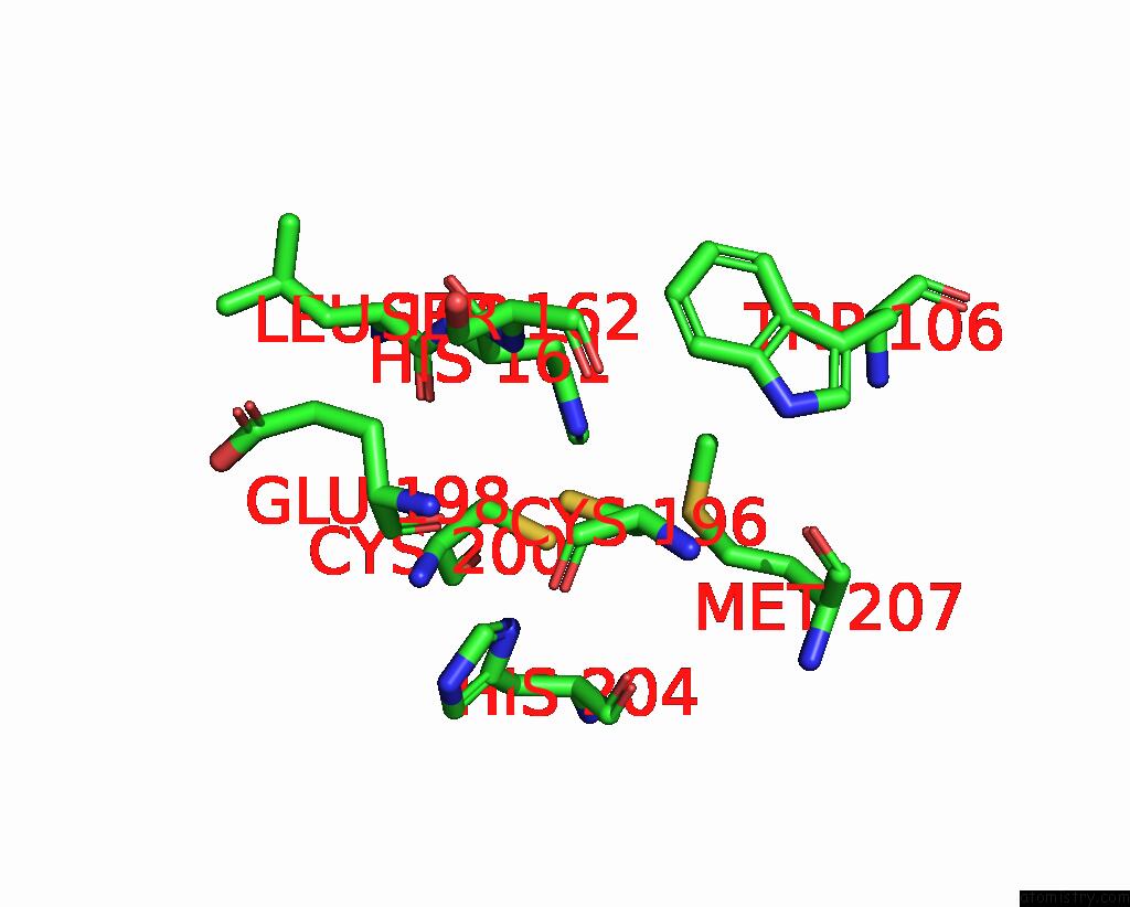
Mono view
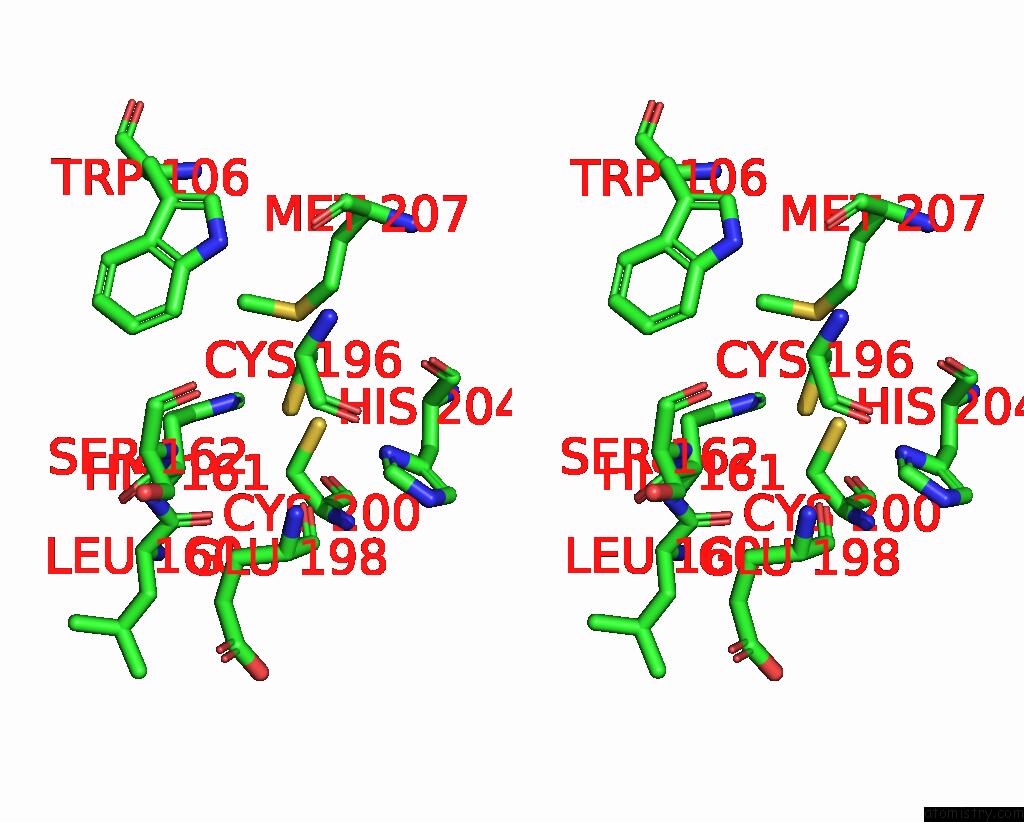
Stereo pair view
|
A full contact list of Copper with other atoms in the Cu binding
site number 5 of Zinc Ion Binding Structure of Bovine Heart Cytochrome C Oxidase in the Fully Oxidized State within 5.0Å range:
probe
|
atom
|
residue
|
distance (Å)
|
B
|
Occ
|
O:Cu228
b:52.2
occ:1.00
|
CU1
|
O:CUA228
|
0.0
|
52.2
|
1.0
|
ND1
|
O:HIS161
|
2.0
|
40.4
|
1.0
|
SG
|
O:CYS196
|
2.2
|
43.4
|
1.0
|
CU2
|
O:CUA228
|
2.3
|
55.7
|
1.0
|
SG
|
O:CYS200
|
2.3
|
43.2
|
1.0
|
SD
|
O:MET207
|
2.9
|
63.6
|
1.0
|
CE1
|
O:HIS161
|
3.0
|
38.8
|
1.0
|
CG
|
O:HIS161
|
3.1
|
43.8
|
1.0
|
CB
|
O:CYS196
|
3.1
|
46.5
|
1.0
|
CE
|
O:MET207
|
3.1
|
48.6
|
1.0
|
CB
|
O:CYS200
|
3.4
|
53.0
|
1.0
|
CB
|
O:HIS161
|
3.5
|
45.6
|
1.0
|
CG
|
O:MET207
|
3.8
|
57.5
|
1.0
|
O
|
O:GLU198
|
3.8
|
54.7
|
1.0
|
CA
|
O:HIS161
|
4.0
|
44.7
|
1.0
|
ND1
|
O:HIS204
|
4.1
|
51.9
|
1.0
|
NE2
|
O:HIS161
|
4.2
|
42.9
|
1.0
|
CD2
|
O:HIS161
|
4.2
|
37.2
|
1.0
|
CA
|
O:CYS196
|
4.5
|
50.3
|
1.0
|
CA
|
O:CYS200
|
4.7
|
55.8
|
1.0
|
O
|
O:LEU160
|
4.7
|
50.5
|
1.0
|
N
|
O:SER162
|
4.8
|
49.2
|
1.0
|
N
|
O:CYS200
|
4.9
|
57.1
|
1.0
|
CA
|
O:HIS204
|
4.9
|
56.6
|
1.0
|
CZ2
|
O:TRP106
|
4.9
|
77.4
|
1.0
|
C
|
O:CYS196
|
4.9
|
53.0
|
1.0
|
O
|
O:HIS204
|
4.9
|
56.5
|
1.0
|
C
|
O:HIS161
|
5.0
|
47.9
|
1.0
|
CE1
|
O:HIS204
|
5.0
|
55.7
|
1.0
|
|
Copper binding site 6 out
of 6 in 2ein
Go back to
Copper Binding Sites List in 2ein
Copper binding site 6 out
of 6 in the Zinc Ion Binding Structure of Bovine Heart Cytochrome C Oxidase in the Fully Oxidized State
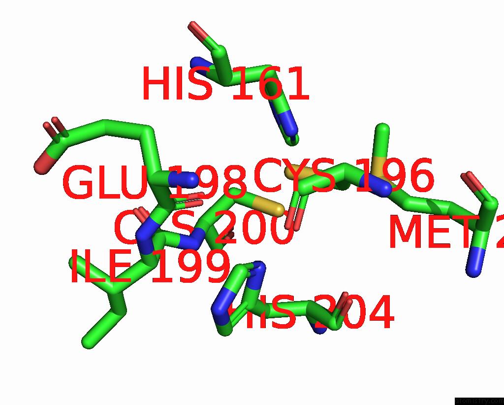
Mono view
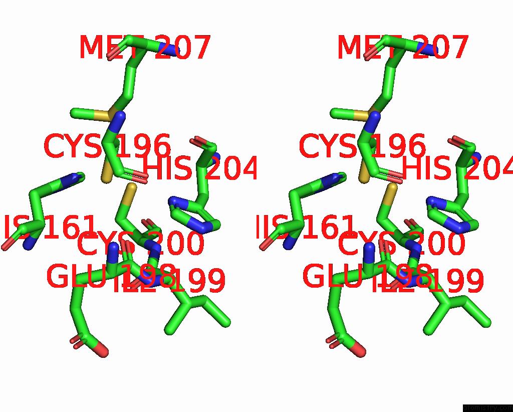
Stereo pair view
|
A full contact list of Copper with other atoms in the Cu binding
site number 6 of Zinc Ion Binding Structure of Bovine Heart Cytochrome C Oxidase in the Fully Oxidized State within 5.0Å range:
probe
|
atom
|
residue
|
distance (Å)
|
B
|
Occ
|
O:Cu228
b:55.7
occ:1.00
|
CU2
|
O:CUA228
|
0.0
|
55.7
|
1.0
|
ND1
|
O:HIS204
|
2.0
|
51.9
|
1.0
|
CU1
|
O:CUA228
|
2.3
|
52.2
|
1.0
|
SG
|
O:CYS200
|
2.3
|
43.2
|
1.0
|
SG
|
O:CYS196
|
2.3
|
43.4
|
1.0
|
O
|
O:GLU198
|
2.4
|
54.7
|
1.0
|
CG
|
O:HIS204
|
2.9
|
54.3
|
1.0
|
CE1
|
O:HIS204
|
3.0
|
55.7
|
1.0
|
CB
|
O:CYS200
|
3.2
|
53.0
|
1.0
|
CB
|
O:HIS204
|
3.3
|
55.0
|
1.0
|
N
|
O:CYS200
|
3.4
|
57.1
|
1.0
|
CB
|
O:CYS196
|
3.5
|
46.5
|
1.0
|
C
|
O:GLU198
|
3.5
|
46.8
|
1.0
|
CA
|
O:HIS204
|
3.5
|
56.6
|
1.0
|
CA
|
O:CYS200
|
3.9
|
55.8
|
1.0
|
ND1
|
O:HIS161
|
3.9
|
40.4
|
1.0
|
O
|
O:HIS204
|
3.9
|
56.5
|
1.0
|
C
|
O:ILE199
|
4.0
|
55.8
|
1.0
|
CD2
|
O:HIS204
|
4.0
|
51.5
|
1.0
|
NE2
|
O:HIS204
|
4.1
|
56.2
|
1.0
|
O
|
O:CYS196
|
4.1
|
53.0
|
1.0
|
C
|
O:HIS204
|
4.2
|
57.8
|
1.0
|
CA
|
O:ILE199
|
4.2
|
54.4
|
1.0
|
N
|
O:ILE199
|
4.3
|
48.0
|
1.0
|
N
|
O:GLU198
|
4.3
|
47.1
|
1.0
|
SD
|
O:MET207
|
4.4
|
63.6
|
1.0
|
C
|
O:CYS196
|
4.4
|
53.0
|
1.0
|
CA
|
O:GLU198
|
4.5
|
44.9
|
1.0
|
CE1
|
O:HIS161
|
4.6
|
38.8
|
1.0
|
CA
|
O:CYS196
|
4.6
|
50.3
|
1.0
|
N
|
O:HIS204
|
4.8
|
58.5
|
1.0
|
O
|
O:ILE199
|
4.8
|
53.4
|
1.0
|
C
|
O:CYS200
|
4.8
|
55.7
|
1.0
|
CG
|
O:MET207
|
4.8
|
57.5
|
1.0
|
CG
|
O:HIS161
|
4.9
|
43.8
|
1.0
|
CA
|
O:HIS161
|
4.9
|
44.7
|
1.0
|
|
Reference:
K.Muramoto,
K.Hirata,
K.Shinzawa-Itoh,
S.Yoko-O,
E.Yamashita,
H.Aoyama,
T.Tsukihara,
S.Yoshikawa.
A Histidine Residue Acting As A Controlling Site For Dioxygen Reduction and Proton Pumping By Cytochrome C Oxidase Proc.Natl.Acad.Sci.Usa V. 104 7881 2007.
ISSN: ISSN 0027-8424
PubMed: 17470809
DOI: 10.1073/PNAS.0610031104
Page generated: Tue Jul 30 23:27:04 2024
|