Copper »
PDB 2cj3-2foy »
2eic »
Copper in PDB 2eic: Crystal Structure of Galactose Oxidase Mutant W290F
Enzymatic activity of Crystal Structure of Galactose Oxidase Mutant W290F
All present enzymatic activity of Crystal Structure of Galactose Oxidase Mutant W290F:
1.1.3.9;
1.1.3.9;
Protein crystallography data
The structure of Crystal Structure of Galactose Oxidase Mutant W290F, PDB code: 2eic
was solved by
N.Akyumani,
S.Tamber,
S.J.Firbank,
P.F.Knowles,
S.E.Phillips,
M.J.Mcpherson,
with X-Ray Crystallography technique. A brief refinement statistics is given in the table below:
| Resolution Low / High (Å) | 30.00 / 2.80 |
| Space group | C 1 2 1 |
| Cell size a, b, c (Å), α, β, γ (°) | 97.770, 88.890, 86.190, 90.00, 117.90, 90.00 |
| R / Rfree (%) | 17.7 / 23.3 |
Other elements in 2eic:
The structure of Crystal Structure of Galactose Oxidase Mutant W290F also contains other interesting chemical elements:
| Sodium | (Na) | 1 atom |
Copper Binding Sites:
The binding sites of Copper atom in the Crystal Structure of Galactose Oxidase Mutant W290F
(pdb code 2eic). This binding sites where shown within
5.0 Angstroms radius around Copper atom.
In total only one binding site of Copper was determined in the Crystal Structure of Galactose Oxidase Mutant W290F, PDB code: 2eic:
In total only one binding site of Copper was determined in the Crystal Structure of Galactose Oxidase Mutant W290F, PDB code: 2eic:
Copper binding site 1 out of 1 in 2eic
Go back to
Copper binding site 1 out
of 1 in the Crystal Structure of Galactose Oxidase Mutant W290F
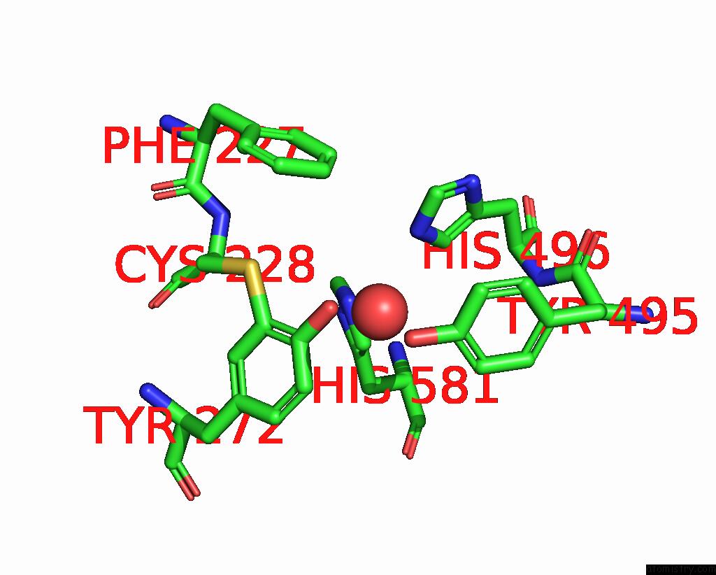
Mono view
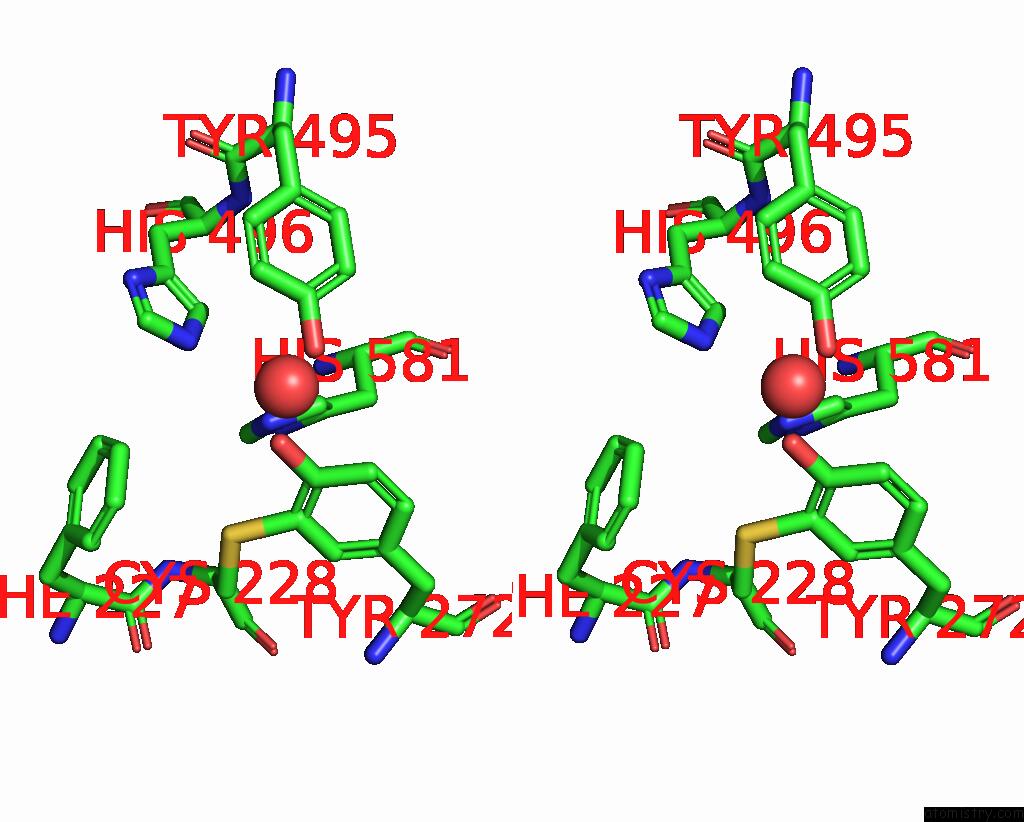
Stereo pair view
Mono view
Stereo pair view
A full contact list of Copper with other atoms in the Cu binding
site number 1 of Crystal Structure of Galactose Oxidase Mutant W290F within 5.0Å range:
|
Reference:
M.S.Rogers,
E.M.Tyler,
N.Akyumani,
C.R.Kurtis,
R.K.Spooner,
S.E.Deacon,
S.Tamber,
S.J.Firbank,
K.Mahmoud,
P.F.Knowles,
S.E.Phillips,
M.J.Mcpherson,
D.M.Dooley.
The Stacking Tryptophan of Galactose Oxidase: A Second-Coordination Sphere Residue That Has Profound Effects on Tyrosyl Radical Behavior and Enzyme Catalysis Biochemistry V. 46 4606 2007.
ISSN: ISSN 0006-2960
PubMed: 17385891
DOI: 10.1021/BI062139D
Page generated: Mon Jul 14 00:56:50 2025
ISSN: ISSN 0006-2960
PubMed: 17385891
DOI: 10.1021/BI062139D
Last articles
Fe in 2B3XFe in 2B11
Fe in 2B12
Fe in 2B10
Fe in 2AYS
Fe in 2B0Z
Fe in 2AZQ
Fe in 2AXT
Fe in 2AV8
Fe in 2AWY