Copper »
PDB 1x9r-2ahl »
2ahk »
Copper in PDB 2ahk: Crystal Structure of the Met-Form of the Copper-Bound Streptomyces Castaneoglobisporus Tyrosinase in Complex with A Caddie Protein Obtained By Soking in Cupric Sulfate For 6 Months
Enzymatic activity of Crystal Structure of the Met-Form of the Copper-Bound Streptomyces Castaneoglobisporus Tyrosinase in Complex with A Caddie Protein Obtained By Soking in Cupric Sulfate For 6 Months
All present enzymatic activity of Crystal Structure of the Met-Form of the Copper-Bound Streptomyces Castaneoglobisporus Tyrosinase in Complex with A Caddie Protein Obtained By Soking in Cupric Sulfate For 6 Months:
1.14.18.1;
1.14.18.1;
Protein crystallography data
The structure of Crystal Structure of the Met-Form of the Copper-Bound Streptomyces Castaneoglobisporus Tyrosinase in Complex with A Caddie Protein Obtained By Soking in Cupric Sulfate For 6 Months, PDB code: 2ahk
was solved by
Y.Matoba,
T.Kumagai,
A.Yamamoto,
H.Yoshitsu,
M.Sugiyama,
with X-Ray Crystallography technique. A brief refinement statistics is given in the table below:
| Resolution Low / High (Å) | 30.00 / 1.71 |
| Space group | P 21 21 2 |
| Cell size a, b, c (Å), α, β, γ (°) | 65.100, 97.470, 54.830, 90.00, 90.00, 90.00 |
| R / Rfree (%) | 19.4 / 24.1 |
Copper Binding Sites:
The binding sites of Copper atom in the Crystal Structure of the Met-Form of the Copper-Bound Streptomyces Castaneoglobisporus Tyrosinase in Complex with A Caddie Protein Obtained By Soking in Cupric Sulfate For 6 Months
(pdb code 2ahk). This binding sites where shown within
5.0 Angstroms radius around Copper atom.
In total 3 binding sites of Copper where determined in the Crystal Structure of the Met-Form of the Copper-Bound Streptomyces Castaneoglobisporus Tyrosinase in Complex with A Caddie Protein Obtained By Soking in Cupric Sulfate For 6 Months, PDB code: 2ahk:
Jump to Copper binding site number: 1; 2; 3;
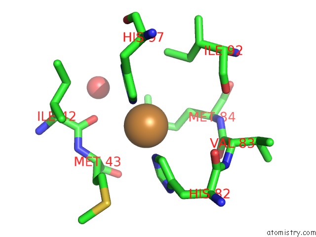
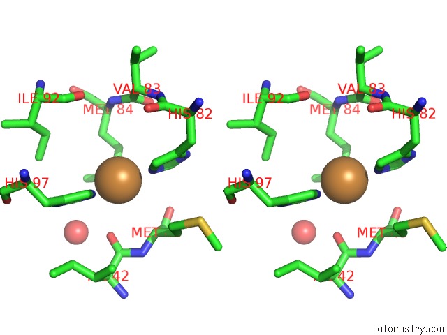
Copper binding site 1 out of 3 in 2ahk
Go back to
Copper binding site 1 out
of 3 in the Crystal Structure of the Met-Form of the Copper-Bound Streptomyces Castaneoglobisporus Tyrosinase in Complex with A Caddie Protein Obtained By Soking in Cupric Sulfate For 6 Months
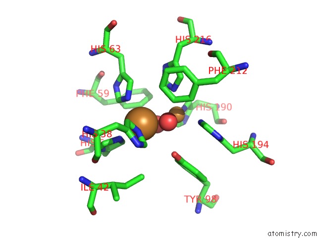
Mono view
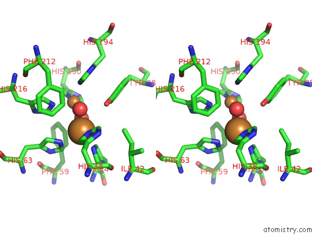
Stereo pair view
Mono view
Stereo pair view
A full contact list of Copper with other atoms in the Cu binding
site number 1 of Crystal Structure of the Met-Form of the Copper-Bound Streptomyces Castaneoglobisporus Tyrosinase in Complex with A Caddie Protein Obtained By Soking in Cupric Sulfate For 6 Months within 5.0Å range:
|
Copper binding site 2 out of 3 in 2ahk
Go back to
Copper binding site 2 out
of 3 in the Crystal Structure of the Met-Form of the Copper-Bound Streptomyces Castaneoglobisporus Tyrosinase in Complex with A Caddie Protein Obtained By Soking in Cupric Sulfate For 6 Months
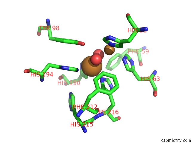
Mono view
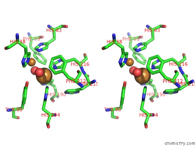
Stereo pair view
Mono view
Stereo pair view
A full contact list of Copper with other atoms in the Cu binding
site number 2 of Crystal Structure of the Met-Form of the Copper-Bound Streptomyces Castaneoglobisporus Tyrosinase in Complex with A Caddie Protein Obtained By Soking in Cupric Sulfate For 6 Months within 5.0Å range:
|
Copper binding site 3 out of 3 in 2ahk
Go back to
Copper binding site 3 out
of 3 in the Crystal Structure of the Met-Form of the Copper-Bound Streptomyces Castaneoglobisporus Tyrosinase in Complex with A Caddie Protein Obtained By Soking in Cupric Sulfate For 6 Months
Mono view
Stereo pair view
Mono view
Stereo pair view
A full contact list of Copper with other atoms in the Cu binding
site number 3 of Crystal Structure of the Met-Form of the Copper-Bound Streptomyces Castaneoglobisporus Tyrosinase in Complex with A Caddie Protein Obtained By Soking in Cupric Sulfate For 6 Months within 5.0Å range:
|
Reference:
Y.Matoba,
T.Kumagai,
A.Yamamoto,
H.Yoshitsu,
M.Sugiyama.
Crystallographic Evidence That the Dinuclear Copper Center of Tyrosinase Is Flexible During Catalysis J.Biol.Chem. V. 281 8981 2006.
ISSN: ISSN 0021-9258
PubMed: 16436386
DOI: 10.1074/JBC.M509785200
Page generated: Tue Jul 30 23:08:05 2024
ISSN: ISSN 0021-9258
PubMed: 16436386
DOI: 10.1074/JBC.M509785200
Last articles
Cl in 4XK2Cl in 4XLG
Cl in 4XLI
Cl in 4XJS
Cl in 4XJI
Cl in 4XJV
Cl in 4XJO
Cl in 4XJD
Cl in 4XJH
Cl in 4XJB