Copper »
PDB 1x9r-2ahl »
2aan »
Copper in PDB 2aan: Auracyanin A: A "Blue" Copper Protein From the Green Thermophilic Photosynthetic Bacterium,Chloroflexus Aurantiacus
Protein crystallography data
The structure of Auracyanin A: A "Blue" Copper Protein From the Green Thermophilic Photosynthetic Bacterium,Chloroflexus Aurantiacus, PDB code: 2aan
was solved by
M.Lee,
J.M.Guss,
H.C.Freeman,
with X-Ray Crystallography technique. A brief refinement statistics is given in the table below:
| Resolution Low / High (Å) | 18.00 / 1.85 |
| Space group | P 41 21 2 |
| Cell size a, b, c (Å), α, β, γ (°) | 70.578, 70.578, 45.665, 90.00, 90.00, 90.00 |
| R / Rfree (%) | 16.2 / 20.7 |
Copper Binding Sites:
The binding sites of Copper atom in the Auracyanin A: A "Blue" Copper Protein From the Green Thermophilic Photosynthetic Bacterium,Chloroflexus Aurantiacus
(pdb code 2aan). This binding sites where shown within
5.0 Angstroms radius around Copper atom.
In total only one binding site of Copper was determined in the Auracyanin A: A "Blue" Copper Protein From the Green Thermophilic Photosynthetic Bacterium,Chloroflexus Aurantiacus, PDB code: 2aan:
In total only one binding site of Copper was determined in the Auracyanin A: A "Blue" Copper Protein From the Green Thermophilic Photosynthetic Bacterium,Chloroflexus Aurantiacus, PDB code: 2aan:
Copper binding site 1 out of 1 in 2aan
Go back to
Copper binding site 1 out
of 1 in the Auracyanin A: A "Blue" Copper Protein From the Green Thermophilic Photosynthetic Bacterium,Chloroflexus Aurantiacus
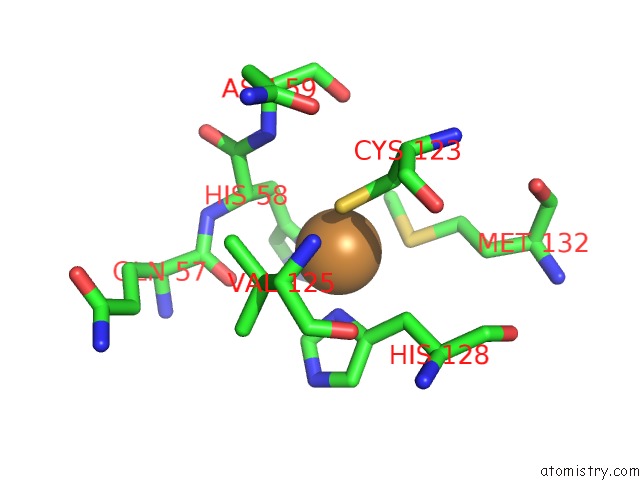
Mono view
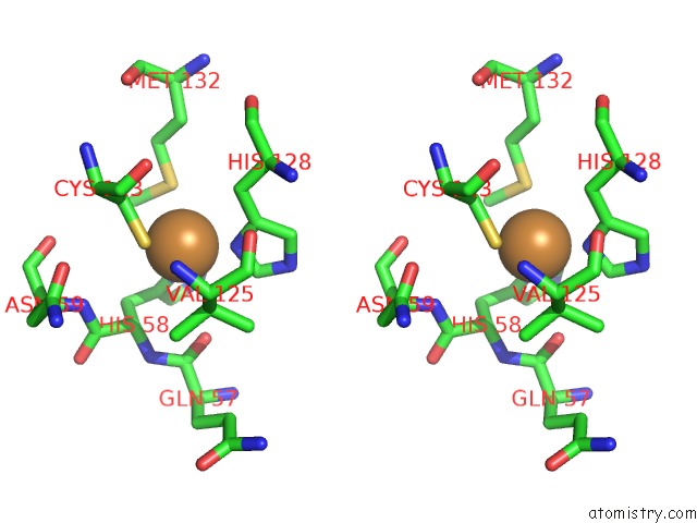
Stereo pair view
Mono view
Stereo pair view
A full contact list of Copper with other atoms in the Cu binding
site number 1 of Auracyanin A: A "Blue" Copper Protein From the Green Thermophilic Photosynthetic Bacterium,Chloroflexus Aurantiacus within 5.0Å range:
|
Reference:
M.Lee,
J.M.Guss,
H.C.Freeman.
The Crystal Structure of Auracyanin A at 1.85 A Resolution: Why the Photosynthetic Bacterium Chloroflexus Needs Two Almost Identical 'Blue' Copper Proteins To Be Published.
Page generated: Tue Jul 30 23:07:24 2024
Last articles
Cl in 5BUFCl in 5BSG
Cl in 5BSF
Cl in 5BSE
Cl in 5BU5
Cl in 5BTV
Cl in 5BT5
Cl in 5BT4
Cl in 5BT3
Cl in 5BRY