Copper »
PDB 1x9r-2ahl »
231d »
Copper in PDB 231d: Structure of A Dna-Porphyrin Complex
Protein crystallography data
The structure of Structure of A Dna-Porphyrin Complex, PDB code: 231d
was solved by
L.A.Lipscomb,
F.X.Zhou,
S.R.Presnell,
R.J.Woo,
M.E.Peek,
R.R.Plaskon,
L.D.Williams,
with X-Ray Crystallography technique. A brief refinement statistics is given in the table below:
| Resolution Low / High (Å) | 8.00 / 2.40 |
| Space group | P 61 2 2 |
| Cell size a, b, c (Å), α, β, γ (°) | 39.490, 39.490, 56.150, 90.00, 90.00, 120.00 |
| R / Rfree (%) | 21.9 / n/a |
Other elements in 231d:
The structure of Structure of A Dna-Porphyrin Complex also contains other interesting chemical elements:
| Sodium | (Na) | 1 atom |
Copper Binding Sites:
The binding sites of Copper atom in the Structure of A Dna-Porphyrin Complex
(pdb code 231d). This binding sites where shown within
5.0 Angstroms radius around Copper atom.
In total only one binding site of Copper was determined in the Structure of A Dna-Porphyrin Complex, PDB code: 231d:
In total only one binding site of Copper was determined in the Structure of A Dna-Porphyrin Complex, PDB code: 231d:
Copper binding site 1 out of 1 in 231d
Go back to
Copper binding site 1 out
of 1 in the Structure of A Dna-Porphyrin Complex

Mono view
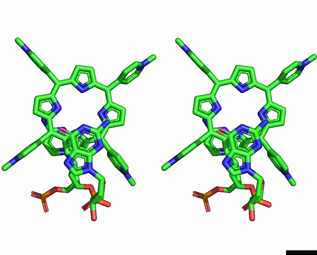
Stereo pair view
Mono view
Stereo pair view
A full contact list of Copper with other atoms in the Cu binding
site number 1 of Structure of A Dna-Porphyrin Complex within 5.0Å range:
|
Reference:
L.A.Lipscomb,
F.X.Zhou,
S.R.Presnell,
R.J.Woo,
M.E.Peek,
R.R.Plaskon,
L.D.Williams.
Structure of Dna-Porphyrin Complex. Biochemistry V. 35 2818 1996.
ISSN: ISSN 0006-2960
PubMed: 8608116
DOI: 10.1021/BI952443Z
Page generated: Mon Jul 14 00:41:48 2025
ISSN: ISSN 0006-2960
PubMed: 8608116
DOI: 10.1021/BI952443Z
Last articles
Fe in 2YXOFe in 2YRS
Fe in 2YXC
Fe in 2YNM
Fe in 2YVJ
Fe in 2YP1
Fe in 2YU2
Fe in 2YU1
Fe in 2YQB
Fe in 2YOO