Copper »
PDB 1x9r-2ahl »
1z9n »
Copper in PDB 1z9n: X-Ray Structure of A Cu-Zn Superoxide Dismutase From Haemophilus Ducreyi with Haem Bound at the Dimer Interface
Enzymatic activity of X-Ray Structure of A Cu-Zn Superoxide Dismutase From Haemophilus Ducreyi with Haem Bound at the Dimer Interface
All present enzymatic activity of X-Ray Structure of A Cu-Zn Superoxide Dismutase From Haemophilus Ducreyi with Haem Bound at the Dimer Interface:
1.15.1.1;
1.15.1.1;
Protein crystallography data
The structure of X-Ray Structure of A Cu-Zn Superoxide Dismutase From Haemophilus Ducreyi with Haem Bound at the Dimer Interface, PDB code: 1z9n
was solved by
K.Djinovic Carugo,
I.Toeroe,
with X-Ray Crystallography technique. A brief refinement statistics is given in the table below:
| Resolution Low / High (Å) | 19.50 / 1.50 |
| Space group | P 1 |
| Cell size a, b, c (Å), α, β, γ (°) | 37.320, 65.900, 69.540, 66.59, 89.87, 76.03 |
| R / Rfree (%) | 15.7 / 19.3 |
Other elements in 1z9n:
The structure of X-Ray Structure of A Cu-Zn Superoxide Dismutase From Haemophilus Ducreyi with Haem Bound at the Dimer Interface also contains other interesting chemical elements:
| Iron | (Fe) | 2 atoms |
| Zinc | (Zn) | 4 atoms |
Copper Binding Sites:
The binding sites of Copper atom in the X-Ray Structure of A Cu-Zn Superoxide Dismutase From Haemophilus Ducreyi with Haem Bound at the Dimer Interface
(pdb code 1z9n). This binding sites where shown within
5.0 Angstroms radius around Copper atom.
In total 4 binding sites of Copper where determined in the X-Ray Structure of A Cu-Zn Superoxide Dismutase From Haemophilus Ducreyi with Haem Bound at the Dimer Interface, PDB code: 1z9n:
Jump to Copper binding site number: 1; 2; 3; 4;
In total 4 binding sites of Copper where determined in the X-Ray Structure of A Cu-Zn Superoxide Dismutase From Haemophilus Ducreyi with Haem Bound at the Dimer Interface, PDB code: 1z9n:
Jump to Copper binding site number: 1; 2; 3; 4;
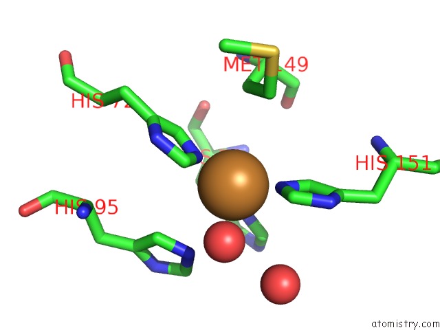
Copper binding site 1 out of 4 in 1z9n
Go back to
Copper binding site 1 out
of 4 in the X-Ray Structure of A Cu-Zn Superoxide Dismutase From Haemophilus Ducreyi with Haem Bound at the Dimer Interface
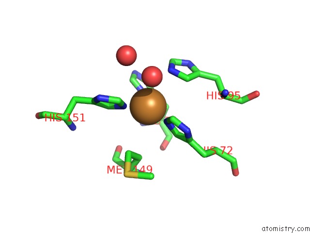
Mono view
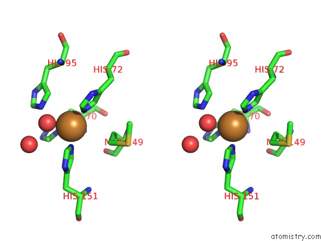
Stereo pair view
Mono view
Stereo pair view
A full contact list of Copper with other atoms in the Cu binding
site number 1 of X-Ray Structure of A Cu-Zn Superoxide Dismutase From Haemophilus Ducreyi with Haem Bound at the Dimer Interface within 5.0Å range:
|
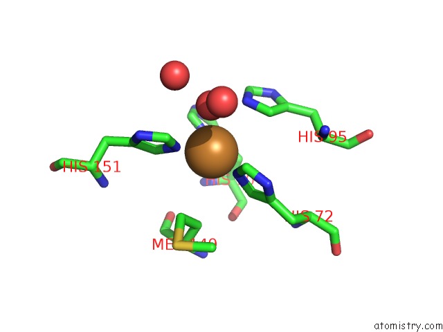
Copper binding site 2 out of 4 in 1z9n
Go back to
Copper binding site 2 out
of 4 in the X-Ray Structure of A Cu-Zn Superoxide Dismutase From Haemophilus Ducreyi with Haem Bound at the Dimer Interface
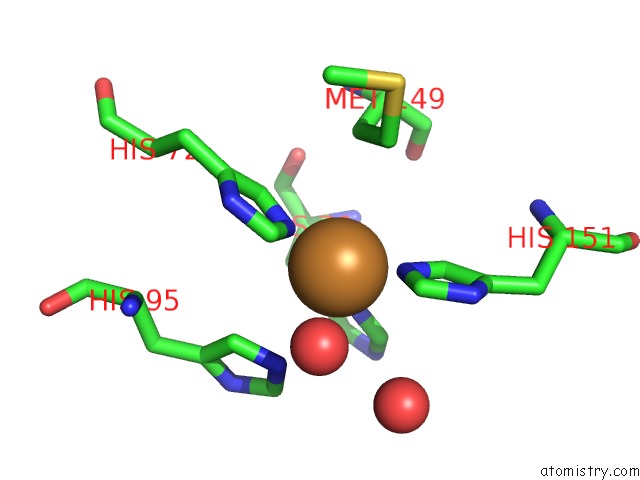
Mono view
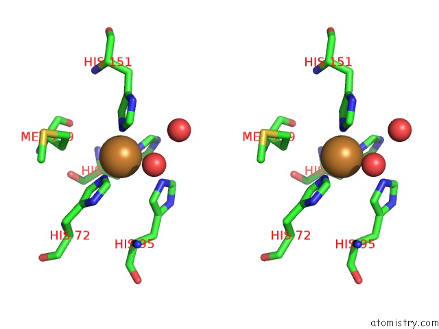
Stereo pair view
Mono view
Stereo pair view
A full contact list of Copper with other atoms in the Cu binding
site number 2 of X-Ray Structure of A Cu-Zn Superoxide Dismutase From Haemophilus Ducreyi with Haem Bound at the Dimer Interface within 5.0Å range:
|
Copper binding site 3 out of 4 in 1z9n
Go back to
Copper binding site 3 out
of 4 in the X-Ray Structure of A Cu-Zn Superoxide Dismutase From Haemophilus Ducreyi with Haem Bound at the Dimer Interface
Mono view

Stereo pair view
Mono view
Stereo pair view
A full contact list of Copper with other atoms in the Cu binding
site number 3 of X-Ray Structure of A Cu-Zn Superoxide Dismutase From Haemophilus Ducreyi with Haem Bound at the Dimer Interface within 5.0Å range:
|
Copper binding site 4 out of 4 in 1z9n
Go back to
Copper binding site 4 out
of 4 in the X-Ray Structure of A Cu-Zn Superoxide Dismutase From Haemophilus Ducreyi with Haem Bound at the Dimer Interface
Mono view
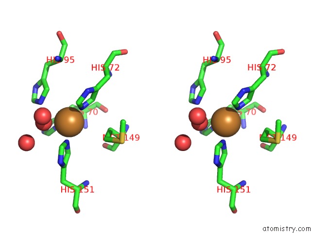
Stereo pair view
Mono view
Stereo pair view
A full contact list of Copper with other atoms in the Cu binding
site number 4 of X-Ray Structure of A Cu-Zn Superoxide Dismutase From Haemophilus Ducreyi with Haem Bound at the Dimer Interface within 5.0Å range:
|
Reference:
I.Toro,
C.Petrutz,
F.Pacello,
M.D'orazio,
A.Battistoni,
K.Djinovic-Carugo.
Structural Basis of Heme Binding in the Cu,Zn Superoxide Dismutase From Haemophilus Ducreyi. J.Mol.Biol. V. 386 406 2009.
ISSN: ISSN 0022-2836
PubMed: 19103206
DOI: 10.1016/J.JMB.2008.12.004
Page generated: Tue Jul 30 23:05:25 2024
ISSN: ISSN 0022-2836
PubMed: 19103206
DOI: 10.1016/J.JMB.2008.12.004
Last articles
Au in 7YJPAu in 7X1R
Au in 7VIU
Au in 7VIT
Au in 7VIS
Au in 7R1M
Au in 7VIR
Au in 7VIQ
Au in 7QVQ
Au in 7VIP