Copper »
PDB 1tmx-1x9l »
1wx2 »
Copper in PDB 1wx2: Crystal Structure of the Oxy-Form of the Copper-Bound Streptomyces Castaneoglobisporus Tyrosinase Complexed with A Caddie Protein Prepared By the Addition of Hydrogenperoxide
Enzymatic activity of Crystal Structure of the Oxy-Form of the Copper-Bound Streptomyces Castaneoglobisporus Tyrosinase Complexed with A Caddie Protein Prepared By the Addition of Hydrogenperoxide
All present enzymatic activity of Crystal Structure of the Oxy-Form of the Copper-Bound Streptomyces Castaneoglobisporus Tyrosinase Complexed with A Caddie Protein Prepared By the Addition of Hydrogenperoxide:
1.14.18.1;
1.14.18.1;
Protein crystallography data
The structure of Crystal Structure of the Oxy-Form of the Copper-Bound Streptomyces Castaneoglobisporus Tyrosinase Complexed with A Caddie Protein Prepared By the Addition of Hydrogenperoxide, PDB code: 1wx2
was solved by
Y.Matoba,
T.Kumagai,
A.Yamamoto,
H.Yoshitsu,
M.Sugiyama,
with X-Ray Crystallography technique. A brief refinement statistics is given in the table below:
| Resolution Low / High (Å) | 30.00 / 1.80 |
| Space group | P 21 21 2 |
| Cell size a, b, c (Å), α, β, γ (°) | 65.160, 96.550, 54.720, 90.00, 90.00, 90.00 |
| R / Rfree (%) | 21 / 24.9 |
Copper Binding Sites:
The binding sites of Copper atom in the Crystal Structure of the Oxy-Form of the Copper-Bound Streptomyces Castaneoglobisporus Tyrosinase Complexed with A Caddie Protein Prepared By the Addition of Hydrogenperoxide
(pdb code 1wx2). This binding sites where shown within
5.0 Angstroms radius around Copper atom.
In total 5 binding sites of Copper where determined in the Crystal Structure of the Oxy-Form of the Copper-Bound Streptomyces Castaneoglobisporus Tyrosinase Complexed with A Caddie Protein Prepared By the Addition of Hydrogenperoxide, PDB code: 1wx2:
Jump to Copper binding site number: 1; 2; 3; 4; 5;
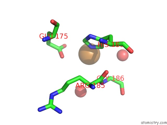
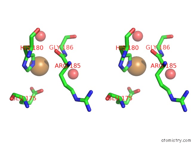
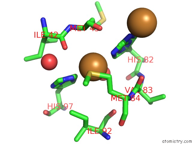
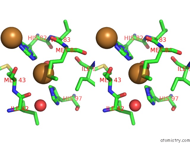
Copper binding site 1 out of 5 in 1wx2
Go back to
Copper binding site 1 out
of 5 in the Crystal Structure of the Oxy-Form of the Copper-Bound Streptomyces Castaneoglobisporus Tyrosinase Complexed with A Caddie Protein Prepared By the Addition of Hydrogenperoxide
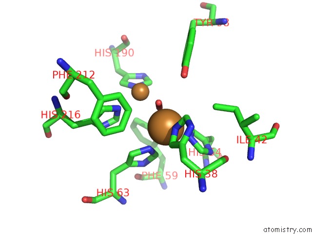
Mono view
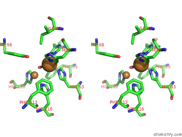
Stereo pair view
Mono view
Stereo pair view
A full contact list of Copper with other atoms in the Cu binding
site number 1 of Crystal Structure of the Oxy-Form of the Copper-Bound Streptomyces Castaneoglobisporus Tyrosinase Complexed with A Caddie Protein Prepared By the Addition of Hydrogenperoxide within 5.0Å range:
|
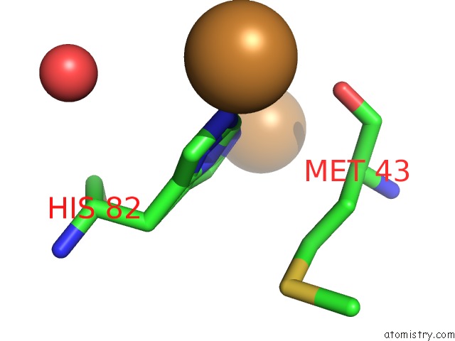
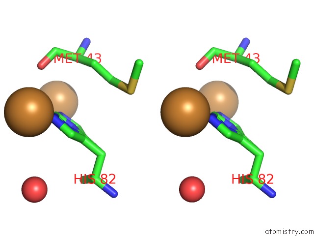
Copper binding site 2 out of 5 in 1wx2
Go back to
Copper binding site 2 out
of 5 in the Crystal Structure of the Oxy-Form of the Copper-Bound Streptomyces Castaneoglobisporus Tyrosinase Complexed with A Caddie Protein Prepared By the Addition of Hydrogenperoxide
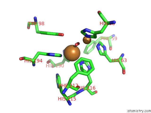
Mono view
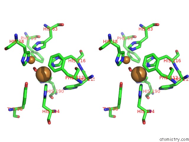
Stereo pair view
Mono view
Stereo pair view
A full contact list of Copper with other atoms in the Cu binding
site number 2 of Crystal Structure of the Oxy-Form of the Copper-Bound Streptomyces Castaneoglobisporus Tyrosinase Complexed with A Caddie Protein Prepared By the Addition of Hydrogenperoxide within 5.0Å range:
|
Copper binding site 3 out of 5 in 1wx2
Go back to
Copper binding site 3 out
of 5 in the Crystal Structure of the Oxy-Form of the Copper-Bound Streptomyces Castaneoglobisporus Tyrosinase Complexed with A Caddie Protein Prepared By the Addition of Hydrogenperoxide
Mono view
Stereo pair view
Mono view
Stereo pair view
A full contact list of Copper with other atoms in the Cu binding
site number 3 of Crystal Structure of the Oxy-Form of the Copper-Bound Streptomyces Castaneoglobisporus Tyrosinase Complexed with A Caddie Protein Prepared By the Addition of Hydrogenperoxide within 5.0Å range:
|
Copper binding site 4 out of 5 in 1wx2
Go back to
Copper binding site 4 out
of 5 in the Crystal Structure of the Oxy-Form of the Copper-Bound Streptomyces Castaneoglobisporus Tyrosinase Complexed with A Caddie Protein Prepared By the Addition of Hydrogenperoxide
Mono view
Stereo pair view
Mono view
Stereo pair view
A full contact list of Copper with other atoms in the Cu binding
site number 4 of Crystal Structure of the Oxy-Form of the Copper-Bound Streptomyces Castaneoglobisporus Tyrosinase Complexed with A Caddie Protein Prepared By the Addition of Hydrogenperoxide within 5.0Å range:
|
Copper binding site 5 out of 5 in 1wx2
Go back to
Copper binding site 5 out
of 5 in the Crystal Structure of the Oxy-Form of the Copper-Bound Streptomyces Castaneoglobisporus Tyrosinase Complexed with A Caddie Protein Prepared By the Addition of Hydrogenperoxide
Mono view
Stereo pair view
Mono view
Stereo pair view
A full contact list of Copper with other atoms in the Cu binding
site number 5 of Crystal Structure of the Oxy-Form of the Copper-Bound Streptomyces Castaneoglobisporus Tyrosinase Complexed with A Caddie Protein Prepared By the Addition of Hydrogenperoxide within 5.0Å range:
|
Reference:
Y.Matoba,
T.Kumagai,
A.Yamamoto,
H.Yoshitsu,
M.Sugiyama.
Crystallographic Evidence That the Dinuclear Copper Center of Tyrosinase Is Flexible During Catalysis J.Biol.Chem. V. 281 8981 2006.
ISSN: ISSN 0021-9258
PubMed: 16436386
DOI: 10.1074/JBC.M509785200
Page generated: Mon Jul 14 00:36:26 2025
ISSN: ISSN 0021-9258
PubMed: 16436386
DOI: 10.1074/JBC.M509785200
Last articles
Fe in 2YXOFe in 2YRS
Fe in 2YXC
Fe in 2YNM
Fe in 2YVJ
Fe in 2YP1
Fe in 2YU2
Fe in 2YU1
Fe in 2YQB
Fe in 2YOO