Copper »
PDB 1tmx-1x9l »
1to5 »
Copper in PDB 1to5: Structure of the Cytosolic Cu,Zn Sod From S. Mansoni
Enzymatic activity of Structure of the Cytosolic Cu,Zn Sod From S. Mansoni
All present enzymatic activity of Structure of the Cytosolic Cu,Zn Sod From S. Mansoni:
1.15.1.1;
1.15.1.1;
Protein crystallography data
The structure of Structure of the Cytosolic Cu,Zn Sod From S. Mansoni, PDB code: 1to5
was solved by
R.M.F.Cardoso,
C.H.T.P.Silva,
A.P.Ulian De Araujo,
T.Tanaka,
M.Tanaka,
R.C.Garratt,
with X-Ray Crystallography technique. A brief refinement statistics is given in the table below:
| Resolution Low / High (Å) | 58.80 / 2.20 |
| Space group | P 21 21 21 |
| Cell size a, b, c (Å), α, β, γ (°) | 74.640, 78.240, 95.180, 90.00, 90.00, 90.00 |
| R / Rfree (%) | 17.6 / 24.1 |
Other elements in 1to5:
The structure of Structure of the Cytosolic Cu,Zn Sod From S. Mansoni also contains other interesting chemical elements:
| Zinc | (Zn) | 4 atoms |
Copper Binding Sites:
The binding sites of Copper atom in the Structure of the Cytosolic Cu,Zn Sod From S. Mansoni
(pdb code 1to5). This binding sites where shown within
5.0 Angstroms radius around Copper atom.
In total 4 binding sites of Copper where determined in the Structure of the Cytosolic Cu,Zn Sod From S. Mansoni, PDB code: 1to5:
Jump to Copper binding site number: 1; 2; 3; 4;
In total 4 binding sites of Copper where determined in the Structure of the Cytosolic Cu,Zn Sod From S. Mansoni, PDB code: 1to5:
Jump to Copper binding site number: 1; 2; 3; 4;
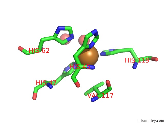
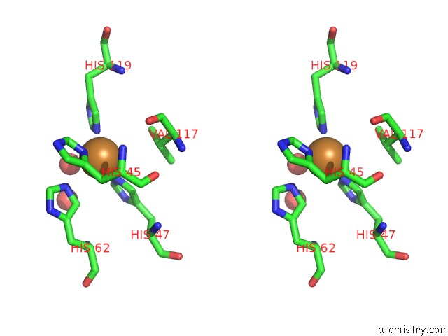
Copper binding site 1 out of 4 in 1to5
Go back to
Copper binding site 1 out
of 4 in the Structure of the Cytosolic Cu,Zn Sod From S. Mansoni
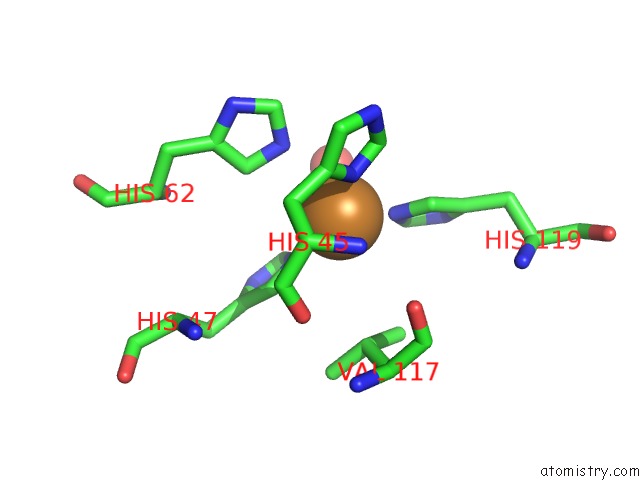
Mono view
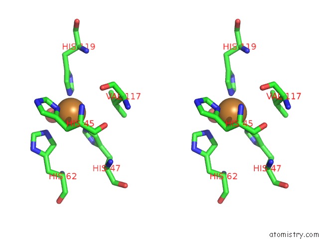
Stereo pair view
Mono view
Stereo pair view
A full contact list of Copper with other atoms in the Cu binding
site number 1 of Structure of the Cytosolic Cu,Zn Sod From S. Mansoni within 5.0Å range:
|
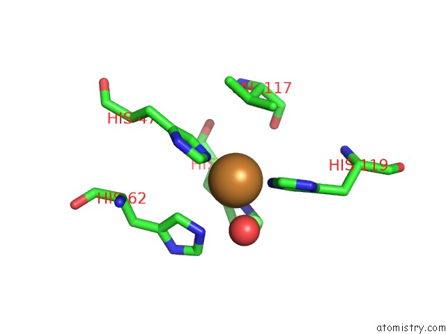
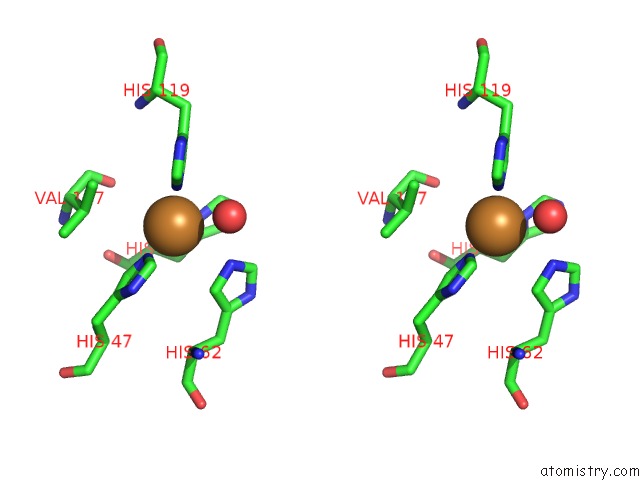
Copper binding site 2 out of 4 in 1to5
Go back to
Copper binding site 2 out
of 4 in the Structure of the Cytosolic Cu,Zn Sod From S. Mansoni
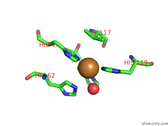
Mono view
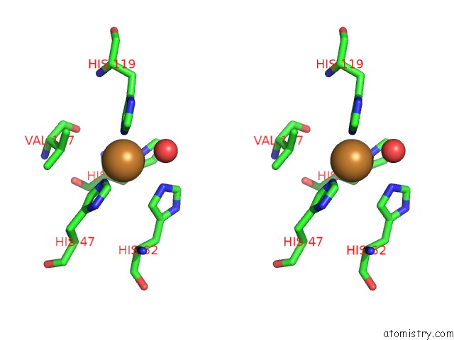
Stereo pair view
Mono view
Stereo pair view
A full contact list of Copper with other atoms in the Cu binding
site number 2 of Structure of the Cytosolic Cu,Zn Sod From S. Mansoni within 5.0Å range:
|
Copper binding site 3 out of 4 in 1to5
Go back to
Copper binding site 3 out
of 4 in the Structure of the Cytosolic Cu,Zn Sod From S. Mansoni
Mono view
Stereo pair view
Mono view
Stereo pair view
A full contact list of Copper with other atoms in the Cu binding
site number 3 of Structure of the Cytosolic Cu,Zn Sod From S. Mansoni within 5.0Å range:
|
Copper binding site 4 out of 4 in 1to5
Go back to
Copper binding site 4 out
of 4 in the Structure of the Cytosolic Cu,Zn Sod From S. Mansoni
Mono view
Stereo pair view
Mono view
Stereo pair view
A full contact list of Copper with other atoms in the Cu binding
site number 4 of Structure of the Cytosolic Cu,Zn Sod From S. Mansoni within 5.0Å range:
|
Reference:
R.M.Cardoso,
C.H.Silva,
A.P.Ulian De Araujo,
T.Tanaka,
M.Tanaka,
R.C.Garratt.
Structure of the Cytosolic Cu,Zn Superoxide Dismutase From Schistosoma Mansoni. Acta Crystallogr.,Sect.D V. 60 1569 2004.
ISSN: ISSN 0907-4449
PubMed: 15333927
DOI: 10.1107/S0907444904016798
Page generated: Mon Jul 14 00:28:58 2025
ISSN: ISSN 0907-4449
PubMed: 15333927
DOI: 10.1107/S0907444904016798
Last articles
Fe in 2YXOFe in 2YRS
Fe in 2YXC
Fe in 2YNM
Fe in 2YVJ
Fe in 2YP1
Fe in 2YU2
Fe in 2YU1
Fe in 2YQB
Fe in 2YOO