Copper »
PDB 1rju-1tl4 »
1tho »
Copper in PDB 1tho: Crystal Structure of A Mutant Escherichia Coli Thioredoxin with An Arginine Insertion in the Active Site
Protein crystallography data
The structure of Crystal Structure of A Mutant Escherichia Coli Thioredoxin with An Arginine Insertion in the Active Site, PDB code: 1tho
was solved by
M.Nikkola,
K.Langsetmo,
J.A.Fuchs,
H.Eklund,
with X-Ray Crystallography technique. A brief refinement statistics is given in the table below:
| Resolution Low / High (Å) | N/A / 2.30 |
| Space group | P 61 |
| Cell size a, b, c (Å), α, β, γ (°) | 78.400, 78.400, 35.100, 90.00, 90.00, 120.00 |
| R / Rfree (%) | 17.3 / n/a |
Copper Binding Sites:
The binding sites of Copper atom in the Crystal Structure of A Mutant Escherichia Coli Thioredoxin with An Arginine Insertion in the Active Site
(pdb code 1tho). This binding sites where shown within
5.0 Angstroms radius around Copper atom.
In total only one binding site of Copper was determined in the Crystal Structure of A Mutant Escherichia Coli Thioredoxin with An Arginine Insertion in the Active Site, PDB code: 1tho:
In total only one binding site of Copper was determined in the Crystal Structure of A Mutant Escherichia Coli Thioredoxin with An Arginine Insertion in the Active Site, PDB code: 1tho:
Copper binding site 1 out of 1 in 1tho
Go back to
Copper binding site 1 out
of 1 in the Crystal Structure of A Mutant Escherichia Coli Thioredoxin with An Arginine Insertion in the Active Site
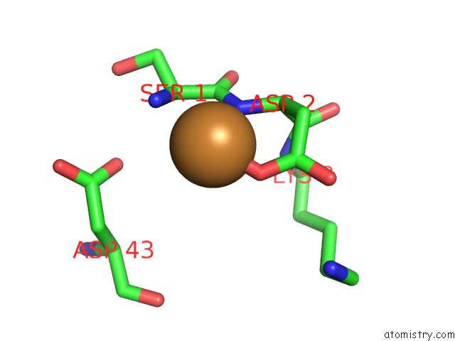
Mono view
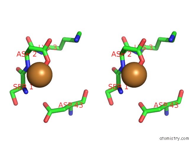
Stereo pair view
Mono view
Stereo pair view
A full contact list of Copper with other atoms in the Cu binding
site number 1 of Crystal Structure of A Mutant Escherichia Coli Thioredoxin with An Arginine Insertion in the Active Site within 5.0Å range:
|
Reference:
M.Nikkola,
K.Langsetmo,
J.A.Fuchs,
H.Eklund.
Crystal Structure of A Mutant Escherichia Coli Thioredoxin with An Arginine Insertion in the Active Site To Be Published.
Page generated: Tue Jul 30 22:50:06 2024
Last articles
Cl in 3F5ECl in 3F5A
Cl in 3F53
Cl in 3F4X
Cl in 3F43
Cl in 3F4M
Cl in 3F4A
Cl in 3F2I
Cl in 3F3S
Cl in 3ETD