Copper »
PDB 1rju-1tl4 »
1slv »
Copper in PDB 1slv: Rat Anionic N143H, E151H Trypsin Complexed to A86H Ecotin; Copper- Bound
Enzymatic activity of Rat Anionic N143H, E151H Trypsin Complexed to A86H Ecotin; Copper- Bound
All present enzymatic activity of Rat Anionic N143H, E151H Trypsin Complexed to A86H Ecotin; Copper- Bound:
3.4.21.4;
3.4.21.4;
Protein crystallography data
The structure of Rat Anionic N143H, E151H Trypsin Complexed to A86H Ecotin; Copper- Bound, PDB code: 1slv
was solved by
L.S.Brinen,
R.J.Fletterick,
with X-Ray Crystallography technique. A brief refinement statistics is given in the table below:
| Resolution Low / High (Å) | 6.00 / 2.30 |
| Space group | C 1 2 1 |
| Cell size a, b, c (Å), α, β, γ (°) | 86.350, 56.590, 81.400, 90.00, 92.99, 90.00 |
| R / Rfree (%) | 19.5 / 27.8 |
Other elements in 1slv:
The structure of Rat Anionic N143H, E151H Trypsin Complexed to A86H Ecotin; Copper- Bound also contains other interesting chemical elements:
| Calcium | (Ca) | 1 atom |
Copper Binding Sites:
The binding sites of Copper atom in the Rat Anionic N143H, E151H Trypsin Complexed to A86H Ecotin; Copper- Bound
(pdb code 1slv). This binding sites where shown within
5.0 Angstroms radius around Copper atom.
In total only one binding site of Copper was determined in the Rat Anionic N143H, E151H Trypsin Complexed to A86H Ecotin; Copper- Bound, PDB code: 1slv:
In total only one binding site of Copper was determined in the Rat Anionic N143H, E151H Trypsin Complexed to A86H Ecotin; Copper- Bound, PDB code: 1slv:
Copper binding site 1 out of 1 in 1slv
Go back to
Copper binding site 1 out
of 1 in the Rat Anionic N143H, E151H Trypsin Complexed to A86H Ecotin; Copper- Bound
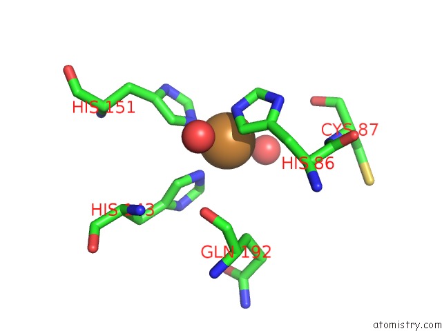
Mono view
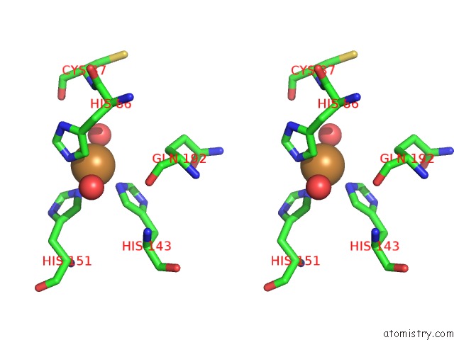
Stereo pair view
Mono view
Stereo pair view
A full contact list of Copper with other atoms in the Cu binding
site number 1 of Rat Anionic N143H, E151H Trypsin Complexed to A86H Ecotin; Copper- Bound within 5.0Å range:
|
Reference:
L.S.Brinen,
W.S.Willett,
C.S.Craik,
R.J.Fletterick.
X-Ray Structures of A Designed Binding Site in Trypsin Show Metal-Dependent Geometry. Biochemistry V. 35 5999 1996.
ISSN: ISSN 0006-2960
PubMed: 8634241
DOI: 10.1021/BI9530200
Page generated: Mon Jul 14 00:25:45 2025
ISSN: ISSN 0006-2960
PubMed: 8634241
DOI: 10.1021/BI9530200
Last articles
Fe in 2YXOFe in 2YRS
Fe in 2YXC
Fe in 2YNM
Fe in 2YVJ
Fe in 2YP1
Fe in 2YU2
Fe in 2YU1
Fe in 2YQB
Fe in 2YOO