Copper »
PDB 1oe2-1rjp »
1qhq »
Copper in PDB 1qhq: Auracyanin, A Blue Copper Protein From the Green Thermophilic Photosynthetic Bacterium Chloroflexus Aurantiacus
Protein crystallography data
The structure of Auracyanin, A Blue Copper Protein From the Green Thermophilic Photosynthetic Bacterium Chloroflexus Aurantiacus, PDB code: 1qhq
was solved by
C.S.Bond,
R.E.Blankenship,
H.C.Freeman,
J.M.Guss,
M.Maher,
F.Selvaraj,
M.C.J.Wilce,
K.Willingham,
with X-Ray Crystallography technique. A brief refinement statistics is given in the table below:
| Resolution Low / High (Å) | 8.00 / 1.55 |
| Space group | P 64 2 2 |
| Cell size a, b, c (Å), α, β, γ (°) | 115.739, 115.739, 54.549, 90.00, 90.00, 120.00 |
| R / Rfree (%) | 19.8 / 23.3 |
Other elements in 1qhq:
The structure of Auracyanin, A Blue Copper Protein From the Green Thermophilic Photosynthetic Bacterium Chloroflexus Aurantiacus also contains other interesting chemical elements:
| Chlorine | (Cl) | 1 atom |
Copper Binding Sites:
The binding sites of Copper atom in the Auracyanin, A Blue Copper Protein From the Green Thermophilic Photosynthetic Bacterium Chloroflexus Aurantiacus
(pdb code 1qhq). This binding sites where shown within
5.0 Angstroms radius around Copper atom.
In total only one binding site of Copper was determined in the Auracyanin, A Blue Copper Protein From the Green Thermophilic Photosynthetic Bacterium Chloroflexus Aurantiacus, PDB code: 1qhq:
In total only one binding site of Copper was determined in the Auracyanin, A Blue Copper Protein From the Green Thermophilic Photosynthetic Bacterium Chloroflexus Aurantiacus, PDB code: 1qhq:
Copper binding site 1 out of 1 in 1qhq
Go back to
Copper binding site 1 out
of 1 in the Auracyanin, A Blue Copper Protein From the Green Thermophilic Photosynthetic Bacterium Chloroflexus Aurantiacus
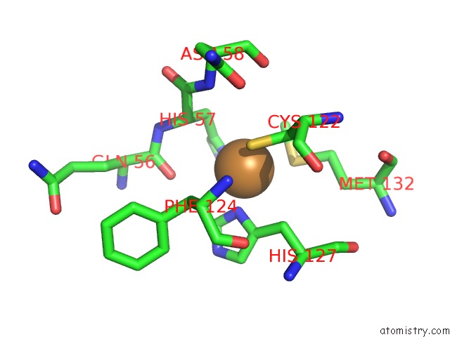
Mono view
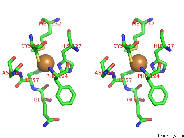
Stereo pair view
Mono view
Stereo pair view
A full contact list of Copper with other atoms in the Cu binding
site number 1 of Auracyanin, A Blue Copper Protein From the Green Thermophilic Photosynthetic Bacterium Chloroflexus Aurantiacus within 5.0Å range:
|
Reference:
C.S.Bond,
R.E.Blankenship,
H.C.Freeman,
J.M.Guss,
M.J.Maher,
F.M.Selvaraj,
M.C.Wilce,
K.M.Willingham.
Crystal Structure of Auracyanin, A "Blue" Copper Protein From the Green Thermophilic Photosynthetic Bacterium Chloroflexus Aurantiacus. J.Mol.Biol. V. 306 47 2001.
ISSN: ISSN 0022-2836
PubMed: 11178893
DOI: 10.1006/JMBI.2000.4201
Page generated: Mon Jul 14 00:18:59 2025
ISSN: ISSN 0022-2836
PubMed: 11178893
DOI: 10.1006/JMBI.2000.4201
Last articles
Fe in 2YXOFe in 2YRS
Fe in 2YXC
Fe in 2YNM
Fe in 2YVJ
Fe in 2YP1
Fe in 2YU2
Fe in 2YU1
Fe in 2YQB
Fe in 2YOO