Copper »
PDB 1oe2-1rjp »
1pnc »
Copper in PDB 1pnc: Accuracy and Precision in Protein Crystal Structure Analysis: Two Independent Refinements of the Structure of Poplar Plastocyanin at 173K
Protein crystallography data
The structure of Accuracy and Precision in Protein Crystal Structure Analysis: Two Independent Refinements of the Structure of Poplar Plastocyanin at 173K, PDB code: 1pnc
was solved by
B.A.Fields,
J.M.Guss,
H.C.Freeman,
with X-Ray Crystallography technique. A brief refinement statistics is given in the table below:
| Resolution Low / High (Å) | 6.00 / 1.60 |
| Space group | P 21 21 21 |
| Cell size a, b, c (Å), α, β, γ (°) | 29.180, 46.290, 56.640, 90.00, 90.00, 90.00 |
| R / Rfree (%) | n/a / n/a |
Copper Binding Sites:
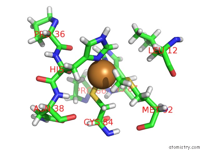
The binding sites of Copper atom in the Accuracy and Precision in Protein Crystal Structure Analysis: Two Independent Refinements of the Structure of Poplar Plastocyanin at 173K
(pdb code 1pnc). This binding sites where shown within
5.0 Angstroms radius around Copper atom.
In total only one binding site of Copper was determined in the Accuracy and Precision in Protein Crystal Structure Analysis: Two Independent Refinements of the Structure of Poplar Plastocyanin at 173K, PDB code: 1pnc:
In total only one binding site of Copper was determined in the Accuracy and Precision in Protein Crystal Structure Analysis: Two Independent Refinements of the Structure of Poplar Plastocyanin at 173K, PDB code: 1pnc:
Copper binding site 1 out of 1 in 1pnc
Go back to
Copper binding site 1 out
of 1 in the Accuracy and Precision in Protein Crystal Structure Analysis: Two Independent Refinements of the Structure of Poplar Plastocyanin at 173K
Mono view
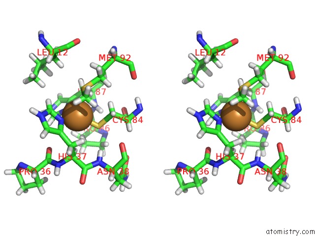
Stereo pair view
Mono view
Stereo pair view
A full contact list of Copper with other atoms in the Cu binding
site number 1 of Accuracy and Precision in Protein Crystal Structure Analysis: Two Independent Refinements of the Structure of Poplar Plastocyanin at 173K within 5.0Å range:
|
Reference:
B.A.Fields,
H.H.Bartsch,
H.D.Bartunik,
F.Cordes,
J.M.Guss,
H.C.Freeman.
Accuracy and Precision in Protein Crystal Structure Analysis: Two Independent Refinements of the Structure of Poplar Plastocyanin at 173 K. Acta Crystallogr.,Sect.D V. 50 709 1994.
ISSN: ISSN 0907-4449
PubMed: 15299368
DOI: 10.1107/S0907444994003021
Page generated: Mon Jul 14 00:17:12 2025
ISSN: ISSN 0907-4449
PubMed: 15299368
DOI: 10.1107/S0907444994003021
Last articles
Fe in 2YXOFe in 2YRS
Fe in 2YXC
Fe in 2YNM
Fe in 2YVJ
Fe in 2YP1
Fe in 2YU2
Fe in 2YU1
Fe in 2YQB
Fe in 2YOO