Copper »
PDB 1oe2-1rjp »
1pcs »
Copper in PDB 1pcs: The 2.15 A Crystal Structure of A Triple Mutant Plastocyanin From the Cyanobacterium Synechocystis Sp. Pcc 6803
Protein crystallography data
The structure of The 2.15 A Crystal Structure of A Triple Mutant Plastocyanin From the Cyanobacterium Synechocystis Sp. Pcc 6803, PDB code: 1pcs
was solved by
A.Romero,
B.De La Cerda,
P.F.Varela,
J.A.Navarro,
M.Hervas,
M.A.De La Rosa,
with X-Ray Crystallography technique. A brief refinement statistics is given in the table below:
| Resolution Low / High (Å) | 8.00 / 2.15 |
| Space group | P 32 2 1 |
| Cell size a, b, c (Å), α, β, γ (°) | 34.300, 34.300, 111.800, 90.00, 90.00, 120.00 |
| R / Rfree (%) | 16.7 / 22 |
Copper Binding Sites:
The binding sites of Copper atom in the The 2.15 A Crystal Structure of A Triple Mutant Plastocyanin From the Cyanobacterium Synechocystis Sp. Pcc 6803
(pdb code 1pcs). This binding sites where shown within
5.0 Angstroms radius around Copper atom.
In total only one binding site of Copper was determined in the The 2.15 A Crystal Structure of A Triple Mutant Plastocyanin From the Cyanobacterium Synechocystis Sp. Pcc 6803, PDB code: 1pcs:
In total only one binding site of Copper was determined in the The 2.15 A Crystal Structure of A Triple Mutant Plastocyanin From the Cyanobacterium Synechocystis Sp. Pcc 6803, PDB code: 1pcs:
Copper binding site 1 out of 1 in 1pcs
Go back to
Copper binding site 1 out
of 1 in the The 2.15 A Crystal Structure of A Triple Mutant Plastocyanin From the Cyanobacterium Synechocystis Sp. Pcc 6803
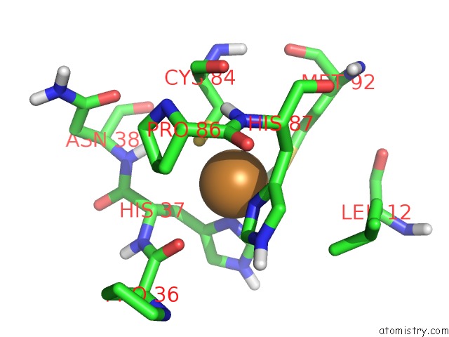
Mono view
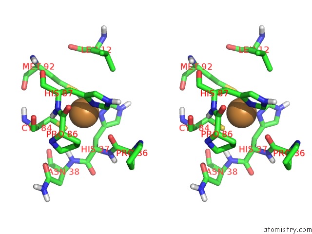
Stereo pair view
Mono view
Stereo pair view
A full contact list of Copper with other atoms in the Cu binding
site number 1 of The 2.15 A Crystal Structure of A Triple Mutant Plastocyanin From the Cyanobacterium Synechocystis Sp. Pcc 6803 within 5.0Å range:
|
Reference:
A.Romero,
B.De La Cerda,
P.F.Varela,
J.A.Navarro,
M.Hervas,
M.A.De La Rosa.
The 2.15 A Crystal Structure of A Triple Mutant Plastocyanin From the Cyanobacterium Synechocystis Sp. Pcc 6803. J.Mol.Biol. V. 275 327 1998.
ISSN: ISSN 0022-2836
PubMed: 9466912
DOI: 10.1006/JMBI.1997.1455
Page generated: Mon Jul 14 00:16:05 2025
ISSN: ISSN 0022-2836
PubMed: 9466912
DOI: 10.1006/JMBI.1997.1455
Last articles
Fe in 2YXOFe in 2YRS
Fe in 2YXC
Fe in 2YNM
Fe in 2YVJ
Fe in 2YP1
Fe in 2YU2
Fe in 2YU1
Fe in 2YQB
Fe in 2YOO