Copper »
PDB 1oe2-1rjp »
1oxy »
Copper in PDB 1oxy: Crystallographic Analysis of Oxygenated and Deoxygenated States of Arthropod Hemocyanin Shows Unusual Differences
Protein crystallography data
The structure of Crystallographic Analysis of Oxygenated and Deoxygenated States of Arthropod Hemocyanin Shows Unusual Differences, PDB code: 1oxy
was solved by
H.Ton-That,
K.Magnus,
with X-Ray Crystallography technique. A brief refinement statistics is given in the table below:
| Resolution Low / High (Å) | 6.00 / 2.40 |
| Space group | H 3 2 |
| Cell size a, b, c (Å), α, β, γ (°) | 117.240, 117.240, 285.860, 90.00, 90.00, 120.00 |
| R / Rfree (%) | 17.1 / n/a |
Copper Binding Sites:
The binding sites of Copper atom in the Crystallographic Analysis of Oxygenated and Deoxygenated States of Arthropod Hemocyanin Shows Unusual Differences
(pdb code 1oxy). This binding sites where shown within
5.0 Angstroms radius around Copper atom.
In total 2 binding sites of Copper where determined in the Crystallographic Analysis of Oxygenated and Deoxygenated States of Arthropod Hemocyanin Shows Unusual Differences, PDB code: 1oxy:
Jump to Copper binding site number: 1; 2;
In total 2 binding sites of Copper where determined in the Crystallographic Analysis of Oxygenated and Deoxygenated States of Arthropod Hemocyanin Shows Unusual Differences, PDB code: 1oxy:
Jump to Copper binding site number: 1; 2;
Copper binding site 1 out of 2 in 1oxy
Go back to
Copper binding site 1 out
of 2 in the Crystallographic Analysis of Oxygenated and Deoxygenated States of Arthropod Hemocyanin Shows Unusual Differences
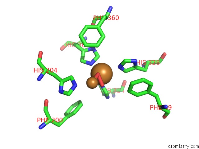
Mono view
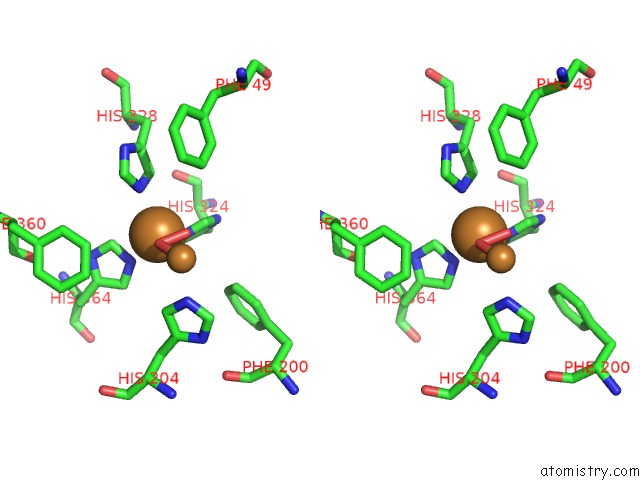
Stereo pair view
Mono view
Stereo pair view
A full contact list of Copper with other atoms in the Cu binding
site number 1 of Crystallographic Analysis of Oxygenated and Deoxygenated States of Arthropod Hemocyanin Shows Unusual Differences within 5.0Å range:
|
Copper binding site 2 out of 2 in 1oxy
Go back to
Copper binding site 2 out
of 2 in the Crystallographic Analysis of Oxygenated and Deoxygenated States of Arthropod Hemocyanin Shows Unusual Differences
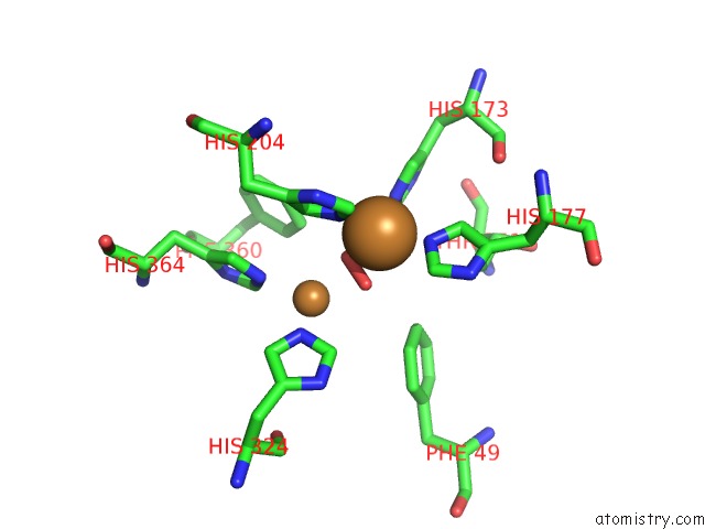
Mono view

Stereo pair view
Mono view
Stereo pair view
A full contact list of Copper with other atoms in the Cu binding
site number 2 of Crystallographic Analysis of Oxygenated and Deoxygenated States of Arthropod Hemocyanin Shows Unusual Differences within 5.0Å range:
|
Reference:
K.A.Magnus,
B.Hazes,
H.Ton-That,
C.Bonaventura,
J.Bonaventura,
W.G.Hol.
Crystallographic Analysis of Oxygenated and Deoxygenated States of Arthropod Hemocyanin Shows Unusual Differences. Proteins V. 19 302 1994.
ISSN: ISSN 0887-3585
PubMed: 7984626
DOI: 10.1002/PROT.340190405
Page generated: Mon Jul 14 00:15:55 2025
ISSN: ISSN 0887-3585
PubMed: 7984626
DOI: 10.1002/PROT.340190405
Last articles
Fe in 2YXOFe in 2YRS
Fe in 2YXC
Fe in 2YNM
Fe in 2YVJ
Fe in 2YP1
Fe in 2YU2
Fe in 2YU1
Fe in 2YQB
Fe in 2YOO