Copper »
PDB 1mg2-1oe1 »
1nol »
Copper in PDB 1nol: Oxygenated Hemocyanin (Subunit Type II)
Protein crystallography data
The structure of Oxygenated Hemocyanin (Subunit Type II), PDB code: 1nol
was solved by
B.Hazes,
W.G.J.Hol,
with X-Ray Crystallography technique. A brief refinement statistics is given in the table below:
| Resolution Low / High (Å) | 8.00 / 2.40 |
| Space group | R 3 2 |
| Cell size a, b, c (Å), α, β, γ (°) | 117.002, 117.002, 117.002, 60.02, 60.02, 60.02 |
| R / Rfree (%) | 18.1 / n/a |
Other elements in 1nol:
The structure of Oxygenated Hemocyanin (Subunit Type II) also contains other interesting chemical elements:
| Calcium | (Ca) | 1 atom |
Copper Binding Sites:
The binding sites of Copper atom in the Oxygenated Hemocyanin (Subunit Type II)
(pdb code 1nol). This binding sites where shown within
5.0 Angstroms radius around Copper atom.
In total 2 binding sites of Copper where determined in the Oxygenated Hemocyanin (Subunit Type II), PDB code: 1nol:
Jump to Copper binding site number: 1; 2;
In total 2 binding sites of Copper where determined in the Oxygenated Hemocyanin (Subunit Type II), PDB code: 1nol:
Jump to Copper binding site number: 1; 2;
Copper binding site 1 out of 2 in 1nol
Go back to
Copper binding site 1 out
of 2 in the Oxygenated Hemocyanin (Subunit Type II)
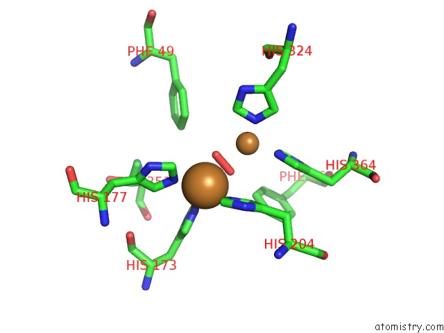
Mono view
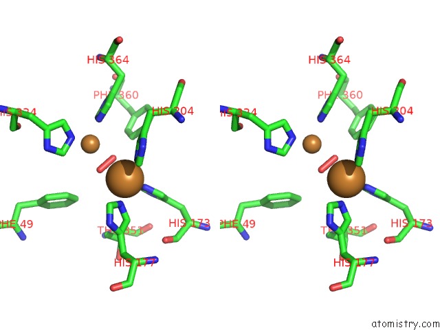
Stereo pair view
Mono view
Stereo pair view
A full contact list of Copper with other atoms in the Cu binding
site number 1 of Oxygenated Hemocyanin (Subunit Type II) within 5.0Å range:
|
Copper binding site 2 out of 2 in 1nol
Go back to
Copper binding site 2 out
of 2 in the Oxygenated Hemocyanin (Subunit Type II)
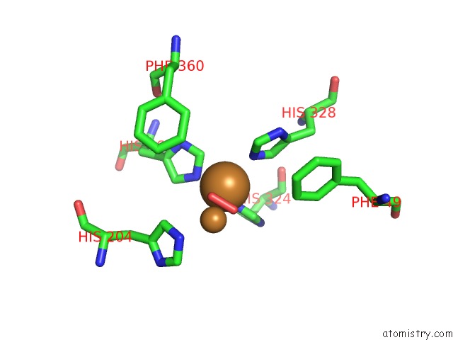
Mono view
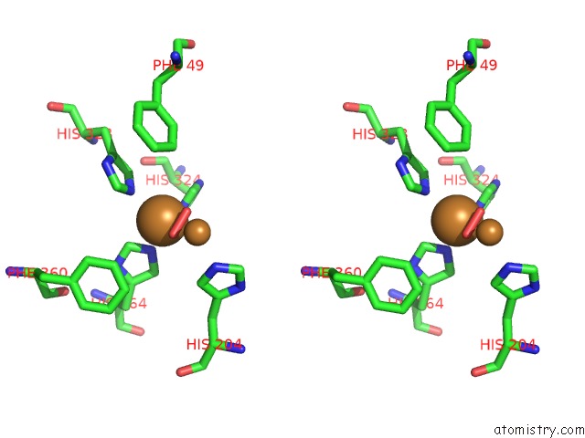
Stereo pair view
Mono view
Stereo pair view
A full contact list of Copper with other atoms in the Cu binding
site number 2 of Oxygenated Hemocyanin (Subunit Type II) within 5.0Å range:
|
Reference:
B.Hazes,
K.A.Magnus,
C.Bonaventura,
J.Bonaventura,
Z.Dauter,
K.H.Kalk,
W.G.Hol.
Crystal Structure of Deoxygenated Limulus Polyphemus Subunit II Hemocyanin at 2.18 A Resolution: Clues For A Mechanism For Allosteric Regulation. Protein Sci. V. 2 597 1993.
ISSN: ISSN 0961-8368
PubMed: 8518732
Page generated: Mon Jul 14 00:11:31 2025
ISSN: ISSN 0961-8368
PubMed: 8518732
Last articles
Fe in 2YXOFe in 2YRS
Fe in 2YXC
Fe in 2YNM
Fe in 2YVJ
Fe in 2YP1
Fe in 2YU2
Fe in 2YU1
Fe in 2YQB
Fe in 2YOO