Copper »
PDB 1jxd-1mfm »
1mfm »
Copper in PDB 1mfm: Monomeric Human Sod Mutant F50E/G51E/E133Q at Atomic Resolution
Enzymatic activity of Monomeric Human Sod Mutant F50E/G51E/E133Q at Atomic Resolution
All present enzymatic activity of Monomeric Human Sod Mutant F50E/G51E/E133Q at Atomic Resolution:
1.15.1.1;
1.15.1.1;
Protein crystallography data
The structure of Monomeric Human Sod Mutant F50E/G51E/E133Q at Atomic Resolution, PDB code: 1mfm
was solved by
M.Ferraroni,
W.Rypniewski,
K.S.Wilson,
P.L.Orioli,
M.S.Viezzoli,
L.Banci,
I.Bertini,
S.Mangani,
with X-Ray Crystallography technique. A brief refinement statistics is given in the table below:
| Resolution Low / High (Å) | 20.00 / 1.02 |
| Space group | P 21 21 21 |
| Cell size a, b, c (Å), α, β, γ (°) | 34.990, 48.110, 81.080, 90.00, 90.00, 90.00 |
| R / Rfree (%) | 11.8 / n/a |
Other elements in 1mfm:
The structure of Monomeric Human Sod Mutant F50E/G51E/E133Q at Atomic Resolution also contains other interesting chemical elements:
| Cadmium | (Cd) | 9 atoms |
| Chlorine | (Cl) | 2 atoms |
| Zinc | (Zn) | 1 atom |
Copper Binding Sites:
The binding sites of Copper atom in the Monomeric Human Sod Mutant F50E/G51E/E133Q at Atomic Resolution
(pdb code 1mfm). This binding sites where shown within
5.0 Angstroms radius around Copper atom.
In total only one binding site of Copper was determined in the Monomeric Human Sod Mutant F50E/G51E/E133Q at Atomic Resolution, PDB code: 1mfm:
In total only one binding site of Copper was determined in the Monomeric Human Sod Mutant F50E/G51E/E133Q at Atomic Resolution, PDB code: 1mfm:
Copper binding site 1 out of 1 in 1mfm
Go back to
Copper binding site 1 out
of 1 in the Monomeric Human Sod Mutant F50E/G51E/E133Q at Atomic Resolution
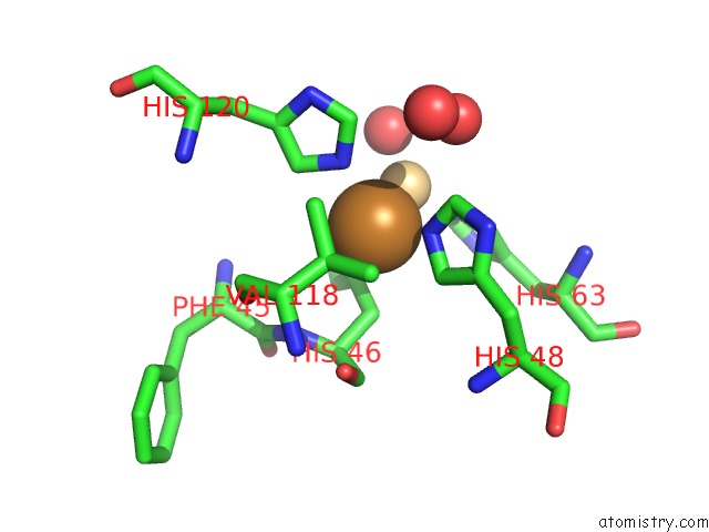
Mono view
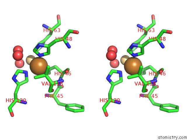
Stereo pair view
Mono view
Stereo pair view
A full contact list of Copper with other atoms in the Cu binding
site number 1 of Monomeric Human Sod Mutant F50E/G51E/E133Q at Atomic Resolution within 5.0Å range:
|
Reference:
M.Ferraroni,
W.Rypniewski,
K.S.Wilson,
M.S.Viezzoli,
L.Banci,
I.Bertini,
S.Mangani.
The Crystal Structure of the Monomeric Human Sod Mutant F50E/G51E/E133Q at Atomic Resolution. the Enzyme Mechanism Revisited. J.Mol.Biol. V. 288 413 1999.
ISSN: ISSN 0022-2836
PubMed: 10329151
DOI: 10.1006/JMBI.1999.2681
Page generated: Tue Jul 30 22:19:58 2024
ISSN: ISSN 0022-2836
PubMed: 10329151
DOI: 10.1006/JMBI.1999.2681
Last articles
Cl in 3GNWCl in 3GNV
Cl in 3GNY
Cl in 3GMF
Cl in 3GNU
Cl in 3GL1
Cl in 3GLM
Cl in 3GL0
Cl in 3GL2
Cl in 3GKY