Copper »
PDB 1jxd-1mfm »
1kyr »
Copper in PDB 1kyr: Crystal Structure of A Cu-Bound Green Fluorescent Protein Zn Biosensor
Protein crystallography data
The structure of Crystal Structure of A Cu-Bound Green Fluorescent Protein Zn Biosensor, PDB code: 1kyr
was solved by
D.P.Barondeau,
C.J.Kassmann,
J.A.Tainer,
E.D.Getzoff,
with X-Ray Crystallography technique. A brief refinement statistics is given in the table below:
| Resolution Low / High (Å) | 20.00 / 1.50 |
| Space group | P 21 21 21 |
| Cell size a, b, c (Å), α, β, γ (°) | 51.166, 62.301, 69.624, 90.00, 90.00, 90.00 |
| R / Rfree (%) | 15 / 21.9 |
Other elements in 1kyr:
The structure of Crystal Structure of A Cu-Bound Green Fluorescent Protein Zn Biosensor also contains other interesting chemical elements:
| Magnesium | (Mg) | 1 atom |
Copper Binding Sites:
The binding sites of Copper atom in the Crystal Structure of A Cu-Bound Green Fluorescent Protein Zn Biosensor
(pdb code 1kyr). This binding sites where shown within
5.0 Angstroms radius around Copper atom.
In total only one binding site of Copper was determined in the Crystal Structure of A Cu-Bound Green Fluorescent Protein Zn Biosensor, PDB code: 1kyr:
In total only one binding site of Copper was determined in the Crystal Structure of A Cu-Bound Green Fluorescent Protein Zn Biosensor, PDB code: 1kyr:
Copper binding site 1 out of 1 in 1kyr
Go back to
Copper binding site 1 out
of 1 in the Crystal Structure of A Cu-Bound Green Fluorescent Protein Zn Biosensor
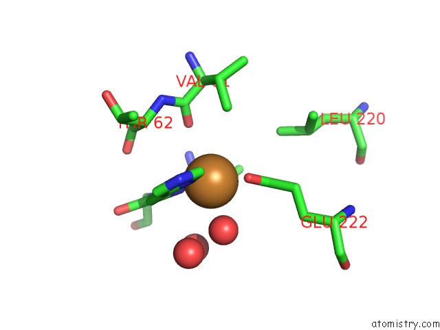
Mono view
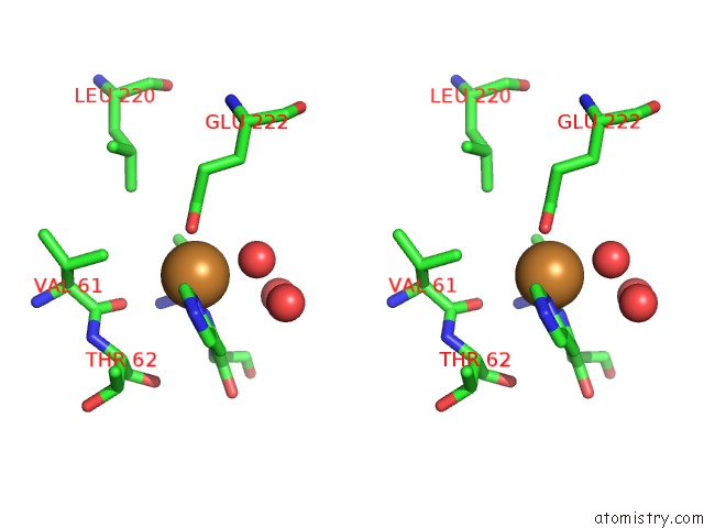
Stereo pair view
Mono view
Stereo pair view
A full contact list of Copper with other atoms in the Cu binding
site number 1 of Crystal Structure of A Cu-Bound Green Fluorescent Protein Zn Biosensor within 5.0Å range:
|
Reference:
D.P.Barondeau,
C.J.Kassmann,
J.A.Tainer,
E.D.Getzoff.
Structural Chemistry of A Green Fluorescent Protein Zn Biosensor. J.Am.Chem.Soc. V. 124 3522 2002.
ISSN: ISSN 0002-7863
PubMed: 11929238
DOI: 10.1021/JA0176954
Page generated: Mon Jul 14 00:00:15 2025
ISSN: ISSN 0002-7863
PubMed: 11929238
DOI: 10.1021/JA0176954
Last articles
Fe in 2YXOFe in 2YRS
Fe in 2YXC
Fe in 2YNM
Fe in 2YVJ
Fe in 2YP1
Fe in 2YU2
Fe in 2YU1
Fe in 2YQB
Fe in 2YOO