Copper »
PDB 1jxd-1mfm »
1keb »
Copper in PDB 1keb: Crystal Structure of Double Mutant M37L,P40S E.Coli Thioredoxin
Protein crystallography data
The structure of Crystal Structure of Double Mutant M37L,P40S E.Coli Thioredoxin, PDB code: 1keb
was solved by
Rudresh,
R.Jain,
V.Dani,
A.Mitra,
S.Srivastava,
S.P.Sarma,
R.Varadarajan,
S.Ramakumar,
with X-Ray Crystallography technique. A brief refinement statistics is given in the table below:
| Resolution Low / High (Å) | 20.00 / 1.80 |
| Space group | P 1 |
| Cell size a, b, c (Å), α, β, γ (°) | 29.320, 37.500, 50.740, 69.25, 79.71, 85.39 |
| R / Rfree (%) | 18.2 / 22.2 |
Copper Binding Sites:
The binding sites of Copper atom in the Crystal Structure of Double Mutant M37L,P40S E.Coli Thioredoxin
(pdb code 1keb). This binding sites where shown within
5.0 Angstroms radius around Copper atom.
In total 2 binding sites of Copper where determined in the Crystal Structure of Double Mutant M37L,P40S E.Coli Thioredoxin, PDB code: 1keb:
Jump to Copper binding site number: 1; 2;
In total 2 binding sites of Copper where determined in the Crystal Structure of Double Mutant M37L,P40S E.Coli Thioredoxin, PDB code: 1keb:
Jump to Copper binding site number: 1; 2;
Copper binding site 1 out of 2 in 1keb
Go back to
Copper binding site 1 out
of 2 in the Crystal Structure of Double Mutant M37L,P40S E.Coli Thioredoxin
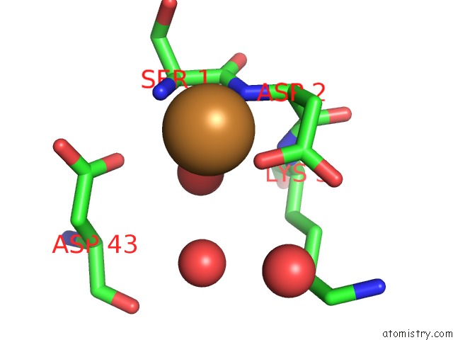
Mono view
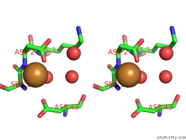
Stereo pair view
Mono view
Stereo pair view
A full contact list of Copper with other atoms in the Cu binding
site number 1 of Crystal Structure of Double Mutant M37L,P40S E.Coli Thioredoxin within 5.0Å range:
|
Copper binding site 2 out of 2 in 1keb
Go back to
Copper binding site 2 out
of 2 in the Crystal Structure of Double Mutant M37L,P40S E.Coli Thioredoxin
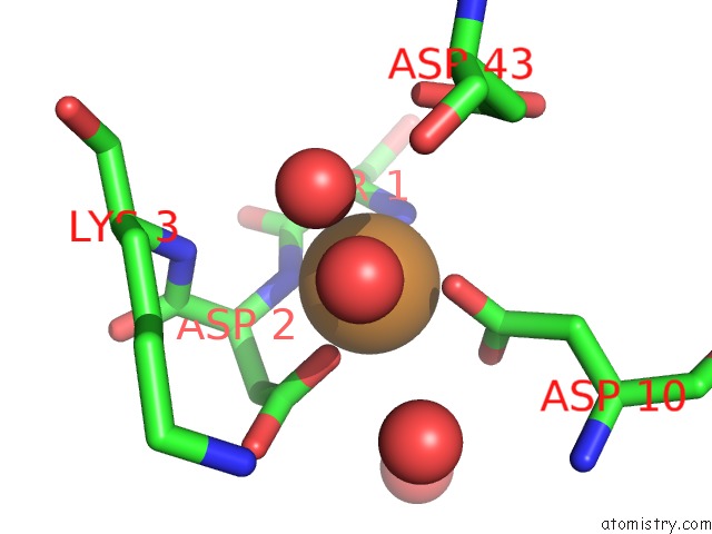
Mono view
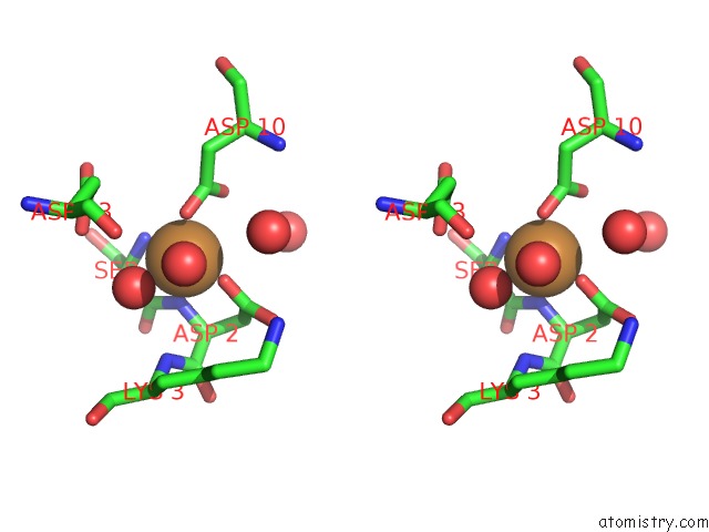
Stereo pair view
Mono view
Stereo pair view
A full contact list of Copper with other atoms in the Cu binding
site number 2 of Crystal Structure of Double Mutant M37L,P40S E.Coli Thioredoxin within 5.0Å range:
|
Reference:
Rudresh,
R.Jain,
V.Dani,
A.Mitra,
S.Srivastava,
S.P.Sarma,
R.Varadarajan,
S.Ramakumar.
Structural Consequences of Replacement of An Alpha-Helical Pro Residue in E.Coli Thioredoxin Protein Eng. V. 15 627 2002.
ISSN: ISSN 0269-2139
PubMed: 12364576
DOI: 10.1093/PROTEIN/15.8.627
Page generated: Tue Jul 30 22:10:49 2024
ISSN: ISSN 0269-2139
PubMed: 12364576
DOI: 10.1093/PROTEIN/15.8.627
Last articles
Cl in 3FAHCl in 3FAK
Cl in 3FA5
Cl in 3F9O
Cl in 3F95
Cl in 3F9P
Cl in 3FA0
Cl in 3F9N
Cl in 3F9L
Cl in 3F8V