Copper »
PDB 1hc1-1jvo »
1ils »
Copper in PDB 1ils: X-Ray Crystal Structure the Two Site-Specific Mutants ILE7SER and PHE110SER of Azurin From Pseudomonas Aeruginosa
Protein crystallography data
The structure of X-Ray Crystal Structure the Two Site-Specific Mutants ILE7SER and PHE110SER of Azurin From Pseudomonas Aeruginosa, PDB code: 1ils
was solved by
C.Hammann,
H.Nar,
R.Huber,
A.Messerschmidt,
with X-Ray Crystallography technique. A brief refinement statistics is given in the table below:
| Resolution Low / High (Å) | 8.00 / 2.20 |
| Space group | P 21 21 21 |
| Cell size a, b, c (Å), α, β, γ (°) | 57.600, 80.700, 110.000, 90.00, 90.00, 90.00 |
| R / Rfree (%) | 16.9 / n/a |
Copper Binding Sites:
The binding sites of Copper atom in the X-Ray Crystal Structure the Two Site-Specific Mutants ILE7SER and PHE110SER of Azurin From Pseudomonas Aeruginosa
(pdb code 1ils). This binding sites where shown within
5.0 Angstroms radius around Copper atom.
In total 4 binding sites of Copper where determined in the X-Ray Crystal Structure the Two Site-Specific Mutants ILE7SER and PHE110SER of Azurin From Pseudomonas Aeruginosa, PDB code: 1ils:
Jump to Copper binding site number: 1; 2; 3; 4;
In total 4 binding sites of Copper where determined in the X-Ray Crystal Structure the Two Site-Specific Mutants ILE7SER and PHE110SER of Azurin From Pseudomonas Aeruginosa, PDB code: 1ils:
Jump to Copper binding site number: 1; 2; 3; 4;
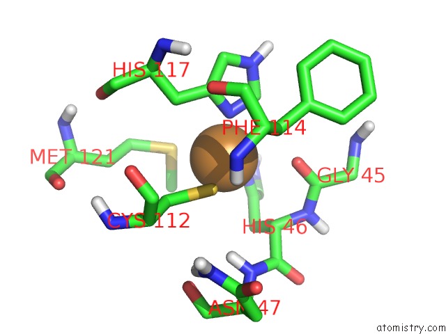
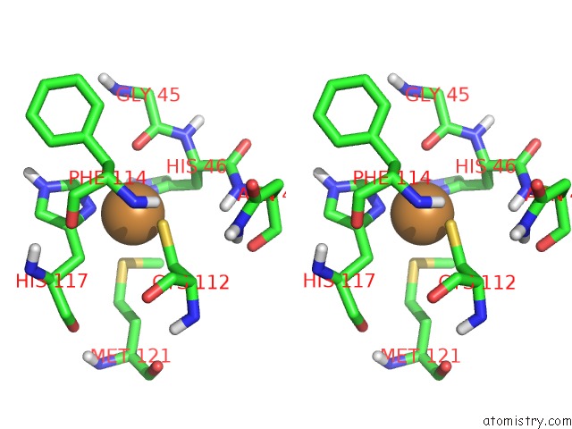
Copper binding site 1 out of 4 in 1ils
Go back to
Copper binding site 1 out
of 4 in the X-Ray Crystal Structure the Two Site-Specific Mutants ILE7SER and PHE110SER of Azurin From Pseudomonas Aeruginosa
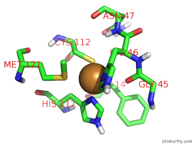
Mono view
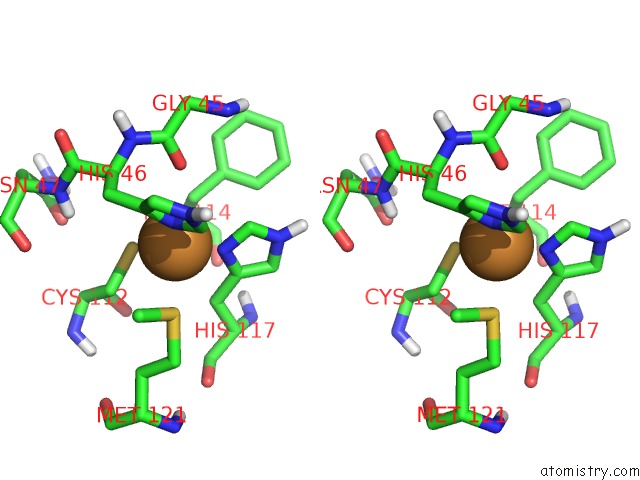
Stereo pair view
Mono view
Stereo pair view
A full contact list of Copper with other atoms in the Cu binding
site number 1 of X-Ray Crystal Structure the Two Site-Specific Mutants ILE7SER and PHE110SER of Azurin From Pseudomonas Aeruginosa within 5.0Å range:
|
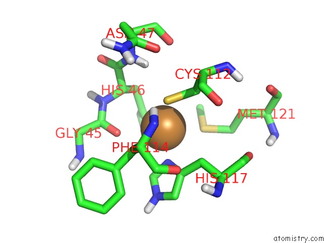
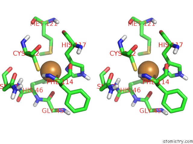
Copper binding site 2 out of 4 in 1ils
Go back to
Copper binding site 2 out
of 4 in the X-Ray Crystal Structure the Two Site-Specific Mutants ILE7SER and PHE110SER of Azurin From Pseudomonas Aeruginosa
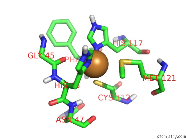
Mono view
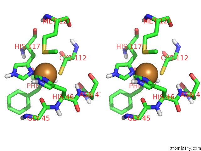
Stereo pair view
Mono view
Stereo pair view
A full contact list of Copper with other atoms in the Cu binding
site number 2 of X-Ray Crystal Structure the Two Site-Specific Mutants ILE7SER and PHE110SER of Azurin From Pseudomonas Aeruginosa within 5.0Å range:
|
Copper binding site 3 out of 4 in 1ils
Go back to
Copper binding site 3 out
of 4 in the X-Ray Crystal Structure the Two Site-Specific Mutants ILE7SER and PHE110SER of Azurin From Pseudomonas Aeruginosa
Mono view
Stereo pair view
Mono view
Stereo pair view
A full contact list of Copper with other atoms in the Cu binding
site number 3 of X-Ray Crystal Structure the Two Site-Specific Mutants ILE7SER and PHE110SER of Azurin From Pseudomonas Aeruginosa within 5.0Å range:
|
Copper binding site 4 out of 4 in 1ils
Go back to
Copper binding site 4 out
of 4 in the X-Ray Crystal Structure the Two Site-Specific Mutants ILE7SER and PHE110SER of Azurin From Pseudomonas Aeruginosa
Mono view
Stereo pair view
Mono view
Stereo pair view
A full contact list of Copper with other atoms in the Cu binding
site number 4 of X-Ray Crystal Structure the Two Site-Specific Mutants ILE7SER and PHE110SER of Azurin From Pseudomonas Aeruginosa within 5.0Å range:
|
Reference:
C.Hammann,
A.Messerschmidt,
R.Huber,
H.Nar,
G.Gilardi,
G.W.Canters.
X-Ray Crystal Structure of the Two Site-Specific Mutants ILE7SER and PHE110SER of Azurin From Pseudomonas Aeruginosa. J.Mol.Biol. V. 255 362 1996.
ISSN: ISSN 0022-2836
PubMed: 8568881
DOI: 10.1006/JMBI.1996.0029
Page generated: Sun Jul 13 23:49:06 2025
ISSN: ISSN 0022-2836
PubMed: 8568881
DOI: 10.1006/JMBI.1996.0029
Last articles
Fe in 2YXOFe in 2YRS
Fe in 2YXC
Fe in 2YNM
Fe in 2YVJ
Fe in 2YP1
Fe in 2YU2
Fe in 2YU1
Fe in 2YQB
Fe in 2YOO