Copper »
PDB 1hc1-1jvo »
1id2 »
Copper in PDB 1id2: Crystal Structure of Amicyanin From Paracoccus Versutus (Thiobacillus Versutus)
Protein crystallography data
The structure of Crystal Structure of Amicyanin From Paracoccus Versutus (Thiobacillus Versutus), PDB code: 1id2
was solved by
A.Romero,
H.Nar,
A.Messerschmidt,
with X-Ray Crystallography technique. A brief refinement statistics is given in the table below:
| Resolution Low / High (Å) | 8.00 / 2.15 |
| Space group | P 32 |
| Cell size a, b, c (Å), α, β, γ (°) | 87.400, 87.400, 38.200, 90.00, 90.00, 120.00 |
| R / Rfree (%) | 17.4 / n/a |
Copper Binding Sites:
The binding sites of Copper atom in the Crystal Structure of Amicyanin From Paracoccus Versutus (Thiobacillus Versutus)
(pdb code 1id2). This binding sites where shown within
5.0 Angstroms radius around Copper atom.
In total 3 binding sites of Copper where determined in the Crystal Structure of Amicyanin From Paracoccus Versutus (Thiobacillus Versutus), PDB code: 1id2:
Jump to Copper binding site number: 1; 2; 3;
In total 3 binding sites of Copper where determined in the Crystal Structure of Amicyanin From Paracoccus Versutus (Thiobacillus Versutus), PDB code: 1id2:
Jump to Copper binding site number: 1; 2; 3;
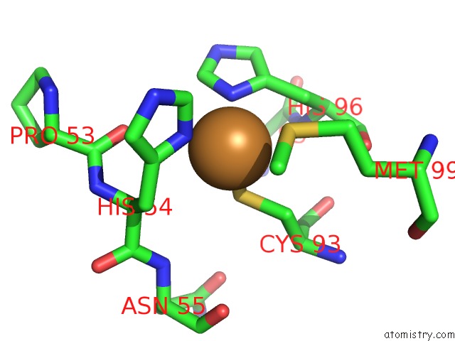
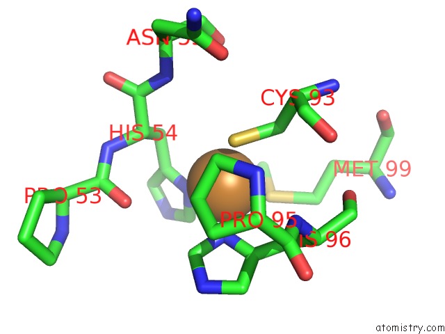
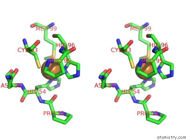
Copper binding site 1 out of 3 in 1id2
Go back to
Copper binding site 1 out
of 3 in the Crystal Structure of Amicyanin From Paracoccus Versutus (Thiobacillus Versutus)
Mono view
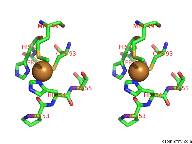
Stereo pair view
Mono view
Stereo pair view
A full contact list of Copper with other atoms in the Cu binding
site number 1 of Crystal Structure of Amicyanin From Paracoccus Versutus (Thiobacillus Versutus) within 5.0Å range:
|
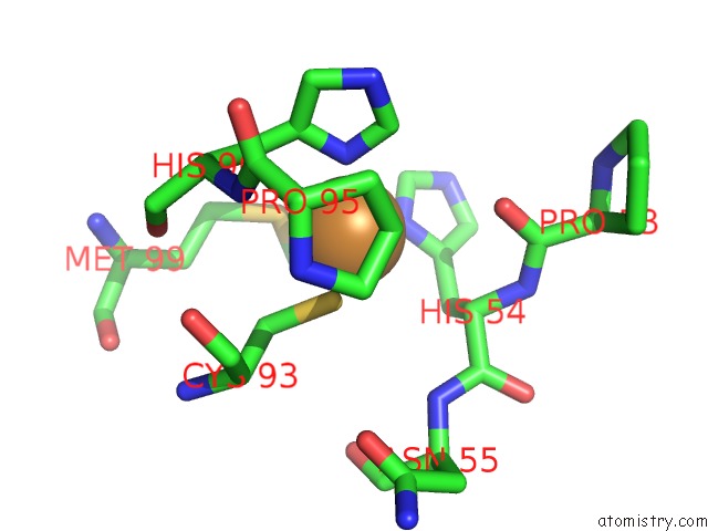
Copper binding site 2 out of 3 in 1id2
Go back to
Copper binding site 2 out
of 3 in the Crystal Structure of Amicyanin From Paracoccus Versutus (Thiobacillus Versutus)
Mono view
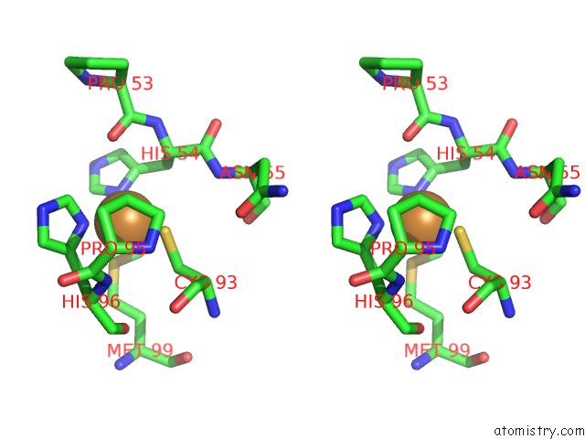
Stereo pair view
Mono view
Stereo pair view
A full contact list of Copper with other atoms in the Cu binding
site number 2 of Crystal Structure of Amicyanin From Paracoccus Versutus (Thiobacillus Versutus) within 5.0Å range:
|
Copper binding site 3 out of 3 in 1id2
Go back to
Copper binding site 3 out
of 3 in the Crystal Structure of Amicyanin From Paracoccus Versutus (Thiobacillus Versutus)
Mono view
Stereo pair view
Mono view
Stereo pair view
A full contact list of Copper with other atoms in the Cu binding
site number 3 of Crystal Structure of Amicyanin From Paracoccus Versutus (Thiobacillus Versutus) within 5.0Å range:
|
Reference:
A.Romero,
H.Nar,
R.Huber,
A.Messerschmidt,
A.P.Kalverda,
G.W.Canters,
R.Durley,
F.S.Mathews.
Crystal Structure Analysis and Refinement at 2.15 A Resolution of Amicyanin, A Type I Blue Copper Protein, From Thiobacillus Versutus. J.Mol.Biol. V. 236 1196 1994.
ISSN: ISSN 0022-2836
PubMed: 8120896
DOI: 10.1016/0022-2836(94)90021-3
Page generated: Sun Jul 13 23:49:03 2025
ISSN: ISSN 0022-2836
PubMed: 8120896
DOI: 10.1016/0022-2836(94)90021-3
Last articles
F in 4C4RF in 4C27
F in 4C1M
F in 4C4D
F in 4C2V
F in 4C28
F in 4C0C
F in 4BYG
F in 4BX2
F in 4BZO