Copper »
PDB 1eso-1haw »
1h1i »
Copper in PDB 1h1i: Crystal Structure of Quercetin 2,3-Dioxygenase Anaerobically Complexed with the Substrate Quercetn
Enzymatic activity of Crystal Structure of Quercetin 2,3-Dioxygenase Anaerobically Complexed with the Substrate Quercetn
All present enzymatic activity of Crystal Structure of Quercetin 2,3-Dioxygenase Anaerobically Complexed with the Substrate Quercetn:
1.13.11.24;
1.13.11.24;
Protein crystallography data
The structure of Crystal Structure of Quercetin 2,3-Dioxygenase Anaerobically Complexed with the Substrate Quercetn, PDB code: 1h1i
was solved by
R.A.Steiner,
B.W.Dijkstra,
with X-Ray Crystallography technique. A brief refinement statistics is given in the table below:
| Resolution Low / High (Å) | 49.39 / 1.75 |
| Space group | P 1 21 1 |
| Cell size a, b, c (Å), α, β, γ (°) | 109.298, 55.745, 124.163, 90.00, 98.39, 90.00 |
| R / Rfree (%) | 14.8 / 18.3 |
Copper Binding Sites:
The binding sites of Copper atom in the Crystal Structure of Quercetin 2,3-Dioxygenase Anaerobically Complexed with the Substrate Quercetn
(pdb code 1h1i). This binding sites where shown within
5.0 Angstroms radius around Copper atom.
In total 4 binding sites of Copper where determined in the Crystal Structure of Quercetin 2,3-Dioxygenase Anaerobically Complexed with the Substrate Quercetn, PDB code: 1h1i:
Jump to Copper binding site number: 1; 2; 3; 4;
In total 4 binding sites of Copper where determined in the Crystal Structure of Quercetin 2,3-Dioxygenase Anaerobically Complexed with the Substrate Quercetn, PDB code: 1h1i:
Jump to Copper binding site number: 1; 2; 3; 4;
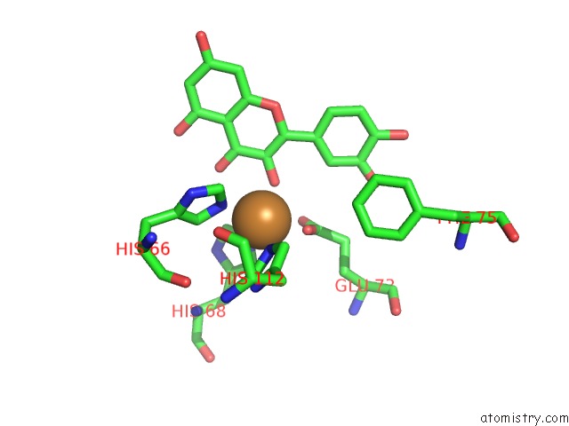
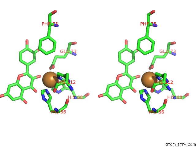
Copper binding site 1 out of 4 in 1h1i
Go back to
Copper binding site 1 out
of 4 in the Crystal Structure of Quercetin 2,3-Dioxygenase Anaerobically Complexed with the Substrate Quercetn
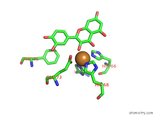
Mono view
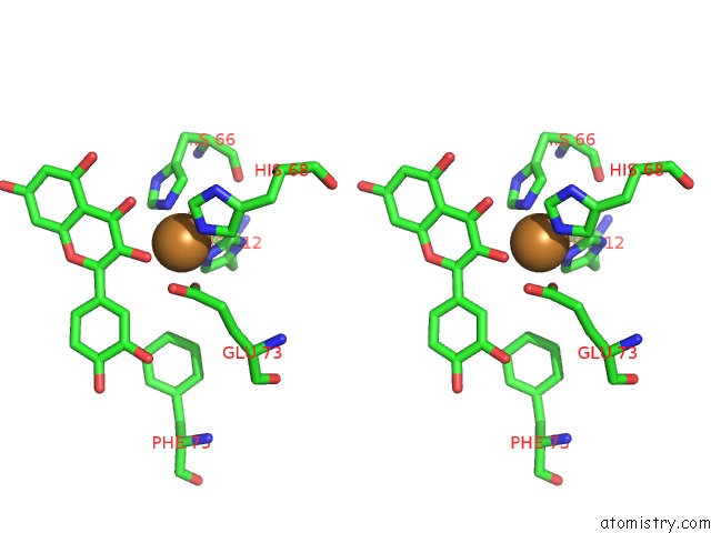
Stereo pair view
Mono view
Stereo pair view
A full contact list of Copper with other atoms in the Cu binding
site number 1 of Crystal Structure of Quercetin 2,3-Dioxygenase Anaerobically Complexed with the Substrate Quercetn within 5.0Å range:
|
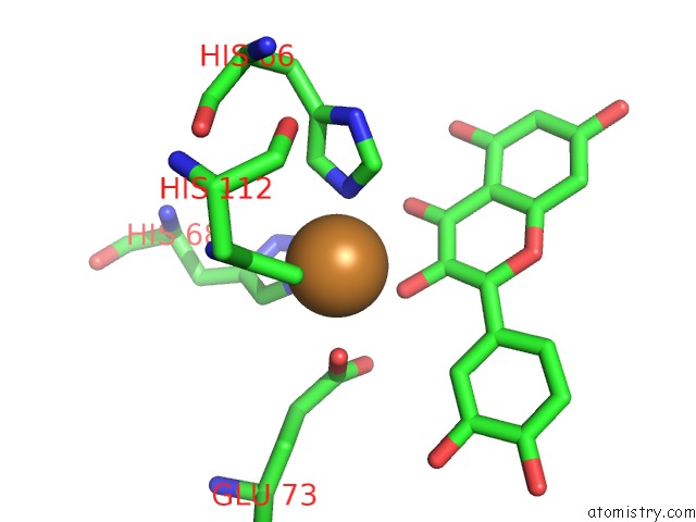
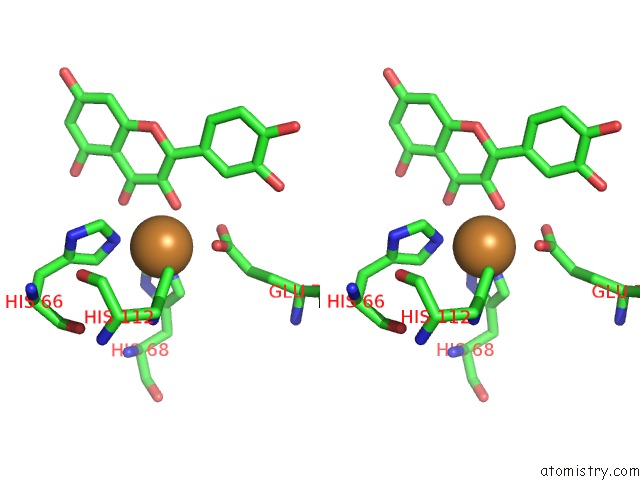
Copper binding site 2 out of 4 in 1h1i
Go back to
Copper binding site 2 out
of 4 in the Crystal Structure of Quercetin 2,3-Dioxygenase Anaerobically Complexed with the Substrate Quercetn
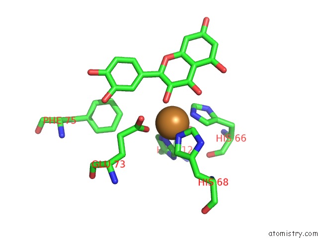
Mono view
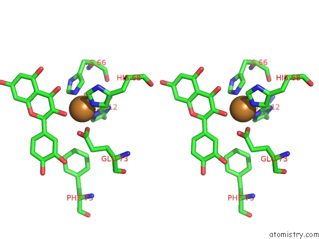
Stereo pair view
Mono view
Stereo pair view
A full contact list of Copper with other atoms in the Cu binding
site number 2 of Crystal Structure of Quercetin 2,3-Dioxygenase Anaerobically Complexed with the Substrate Quercetn within 5.0Å range:
|
Copper binding site 3 out of 4 in 1h1i
Go back to
Copper binding site 3 out
of 4 in the Crystal Structure of Quercetin 2,3-Dioxygenase Anaerobically Complexed with the Substrate Quercetn
Mono view
Stereo pair view
Mono view
Stereo pair view
A full contact list of Copper with other atoms in the Cu binding
site number 3 of Crystal Structure of Quercetin 2,3-Dioxygenase Anaerobically Complexed with the Substrate Quercetn within 5.0Å range:
|
Copper binding site 4 out of 4 in 1h1i
Go back to
Copper binding site 4 out
of 4 in the Crystal Structure of Quercetin 2,3-Dioxygenase Anaerobically Complexed with the Substrate Quercetn
Mono view
Stereo pair view
Mono view
Stereo pair view
A full contact list of Copper with other atoms in the Cu binding
site number 4 of Crystal Structure of Quercetin 2,3-Dioxygenase Anaerobically Complexed with the Substrate Quercetn within 5.0Å range:
|
Reference:
R.A.Steiner,
K.H.Kalk,
B.W.Dijkstra.
Anaerobic Enzyme.Substrate Structures Provide Insight Into the Reaction Mechanism of the Copper- Dependent Quercetin 2,3-Dioxygenase. Proc.Natl.Acad.Sci.Usa V. 99 16625 2002.
ISSN: ISSN 0027-8424
PubMed: 12486225
DOI: 10.1073/PNAS.262506299
Page generated: Tue Jul 30 21:52:44 2024
ISSN: ISSN 0027-8424
PubMed: 12486225
DOI: 10.1073/PNAS.262506299
Last articles
Zn in 9MJ5Zn in 9HNW
Zn in 9G0L
Zn in 9FNE
Zn in 9DZN
Zn in 9E0I
Zn in 9D32
Zn in 9DAK
Zn in 8ZXC
Zn in 8ZUF