Copper »
PDB 1bex-1eqw »
1ekj »
Copper in PDB 1ekj: The X-Ray Crystallographic Structure of Beta Carbonic Anhydrase From the C3 Dicot Pisum Sativum
Enzymatic activity of The X-Ray Crystallographic Structure of Beta Carbonic Anhydrase From the C3 Dicot Pisum Sativum
All present enzymatic activity of The X-Ray Crystallographic Structure of Beta Carbonic Anhydrase From the C3 Dicot Pisum Sativum:
4.2.1.1;
4.2.1.1;
Protein crystallography data
The structure of The X-Ray Crystallographic Structure of Beta Carbonic Anhydrase From the C3 Dicot Pisum Sativum, PDB code: 1ekj
was solved by
M.S.Kimber,
E.F.Pai,
with X-Ray Crystallography technique. A brief refinement statistics is given in the table below:
| Resolution Low / High (Å) | 40.00 / 1.93 |
| Space group | C 2 2 2 |
| Cell size a, b, c (Å), α, β, γ (°) | 136.909, 143.318, 202.135, 90.00, 90.00, 90.00 |
| R / Rfree (%) | 22.9 / 25 |
Other elements in 1ekj:
The structure of The X-Ray Crystallographic Structure of Beta Carbonic Anhydrase From the C3 Dicot Pisum Sativum also contains other interesting chemical elements:
| Chlorine | (Cl) | 8 atoms |
| Zinc | (Zn) | 8 atoms |
Copper Binding Sites:
The binding sites of Copper atom in the The X-Ray Crystallographic Structure of Beta Carbonic Anhydrase From the C3 Dicot Pisum Sativum
(pdb code 1ekj). This binding sites where shown within
5.0 Angstroms radius around Copper atom.
In total 4 binding sites of Copper where determined in the The X-Ray Crystallographic Structure of Beta Carbonic Anhydrase From the C3 Dicot Pisum Sativum, PDB code: 1ekj:
Jump to Copper binding site number: 1; 2; 3; 4;
In total 4 binding sites of Copper where determined in the The X-Ray Crystallographic Structure of Beta Carbonic Anhydrase From the C3 Dicot Pisum Sativum, PDB code: 1ekj:
Jump to Copper binding site number: 1; 2; 3; 4;
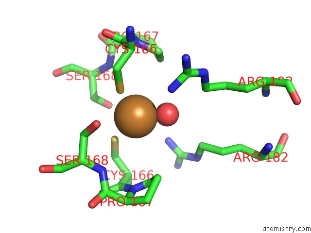
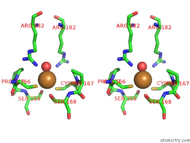
Copper binding site 1 out of 4 in 1ekj
Go back to
Copper binding site 1 out
of 4 in the The X-Ray Crystallographic Structure of Beta Carbonic Anhydrase From the C3 Dicot Pisum Sativum
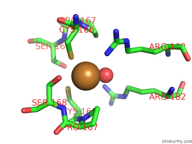
Mono view
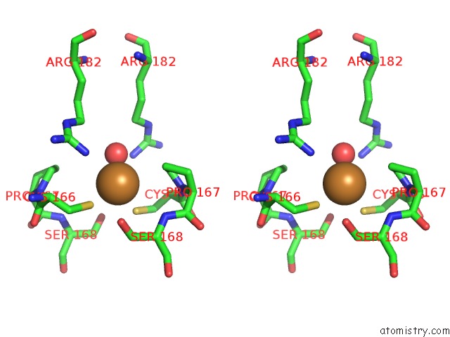
Stereo pair view
Mono view
Stereo pair view
A full contact list of Copper with other atoms in the Cu binding
site number 1 of The X-Ray Crystallographic Structure of Beta Carbonic Anhydrase From the C3 Dicot Pisum Sativum within 5.0Å range:
|
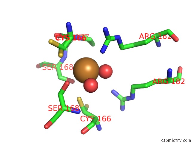
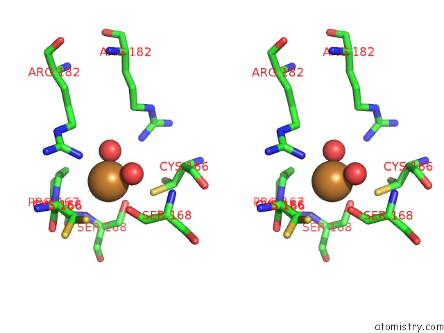
Copper binding site 2 out of 4 in 1ekj
Go back to
Copper binding site 2 out
of 4 in the The X-Ray Crystallographic Structure of Beta Carbonic Anhydrase From the C3 Dicot Pisum Sativum
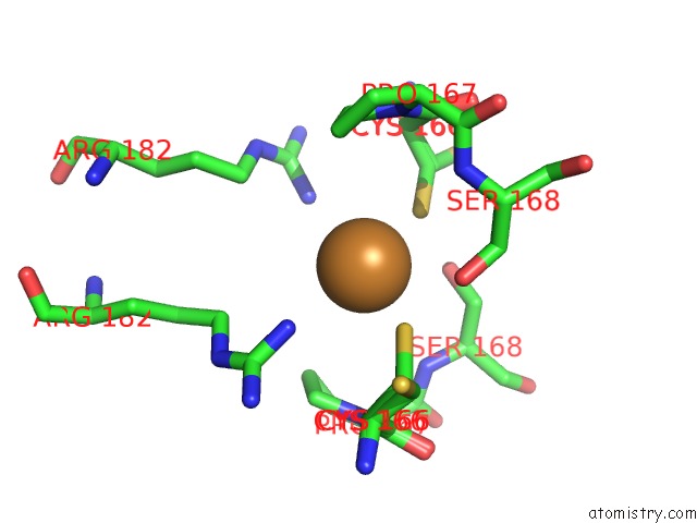
Mono view
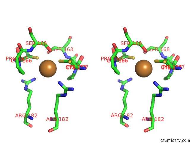
Stereo pair view
Mono view
Stereo pair view
A full contact list of Copper with other atoms in the Cu binding
site number 2 of The X-Ray Crystallographic Structure of Beta Carbonic Anhydrase From the C3 Dicot Pisum Sativum within 5.0Å range:
|
Copper binding site 3 out of 4 in 1ekj
Go back to
Copper binding site 3 out
of 4 in the The X-Ray Crystallographic Structure of Beta Carbonic Anhydrase From the C3 Dicot Pisum Sativum
Mono view
Stereo pair view
Mono view
Stereo pair view
A full contact list of Copper with other atoms in the Cu binding
site number 3 of The X-Ray Crystallographic Structure of Beta Carbonic Anhydrase From the C3 Dicot Pisum Sativum within 5.0Å range:
|
Copper binding site 4 out of 4 in 1ekj
Go back to
Copper binding site 4 out
of 4 in the The X-Ray Crystallographic Structure of Beta Carbonic Anhydrase From the C3 Dicot Pisum Sativum
Mono view
Stereo pair view
Mono view
Stereo pair view
A full contact list of Copper with other atoms in the Cu binding
site number 4 of The X-Ray Crystallographic Structure of Beta Carbonic Anhydrase From the C3 Dicot Pisum Sativum within 5.0Å range:
|
Reference:
M.S.Kimber,
E.F.Pai.
The Active Site Architecture of Pisum Sativum Beta-Carbonic Anhydrase Is A Mirror Image of That of Alpha-Carbonic Anhydrases. Embo J. V. 19 1407 2000.
ISSN: ISSN 0261-4189
PubMed: 10747009
DOI: 10.1093/EMBOJ/19.7.1407
Page generated: Tue Jul 30 21:44:48 2024
ISSN: ISSN 0261-4189
PubMed: 10747009
DOI: 10.1093/EMBOJ/19.7.1407
Last articles
Zn in 9MJ5Zn in 9HNW
Zn in 9G0L
Zn in 9FNE
Zn in 9DZN
Zn in 9E0I
Zn in 9D32
Zn in 9DAK
Zn in 8ZXC
Zn in 8ZUF