Copper »
PDB 1bex-1eqw »
1d6y »
Copper in PDB 1d6y: Crystal Structure of E. Coli Copper-Containing Amine Oxidase Anaerobically Reduced with Beta-Phenylethylamine and Complexed with Nitric Oxide.
Enzymatic activity of Crystal Structure of E. Coli Copper-Containing Amine Oxidase Anaerobically Reduced with Beta-Phenylethylamine and Complexed with Nitric Oxide.
All present enzymatic activity of Crystal Structure of E. Coli Copper-Containing Amine Oxidase Anaerobically Reduced with Beta-Phenylethylamine and Complexed with Nitric Oxide.:
1.4.3.6;
1.4.3.6;
Protein crystallography data
The structure of Crystal Structure of E. Coli Copper-Containing Amine Oxidase Anaerobically Reduced with Beta-Phenylethylamine and Complexed with Nitric Oxide., PDB code: 1d6y
was solved by
C.M.Wilmot,
J.Hajdu,
M.J.Mcpherson,
P.F.Knowles,
S.E.V.Phillips,
with X-Ray Crystallography technique. A brief refinement statistics is given in the table below:
| Resolution Low / High (Å) | 30.00 / 2.40 |
| Space group | P 21 21 21 |
| Cell size a, b, c (Å), α, β, γ (°) | 135.236, 166.482, 79.628, 90.00, 90.00, 90.00 |
| R / Rfree (%) | 18.1 / 23.1 |
Other elements in 1d6y:
The structure of Crystal Structure of E. Coli Copper-Containing Amine Oxidase Anaerobically Reduced with Beta-Phenylethylamine and Complexed with Nitric Oxide. also contains other interesting chemical elements:
| Calcium | (Ca) | 4 atoms |
Copper Binding Sites:
The binding sites of Copper atom in the Crystal Structure of E. Coli Copper-Containing Amine Oxidase Anaerobically Reduced with Beta-Phenylethylamine and Complexed with Nitric Oxide.
(pdb code 1d6y). This binding sites where shown within
5.0 Angstroms radius around Copper atom.
In total 2 binding sites of Copper where determined in the Crystal Structure of E. Coli Copper-Containing Amine Oxidase Anaerobically Reduced with Beta-Phenylethylamine and Complexed with Nitric Oxide., PDB code: 1d6y:
Jump to Copper binding site number: 1; 2;
In total 2 binding sites of Copper where determined in the Crystal Structure of E. Coli Copper-Containing Amine Oxidase Anaerobically Reduced with Beta-Phenylethylamine and Complexed with Nitric Oxide., PDB code: 1d6y:
Jump to Copper binding site number: 1; 2;
Copper binding site 1 out of 2 in 1d6y
Go back to
Copper binding site 1 out
of 2 in the Crystal Structure of E. Coli Copper-Containing Amine Oxidase Anaerobically Reduced with Beta-Phenylethylamine and Complexed with Nitric Oxide.
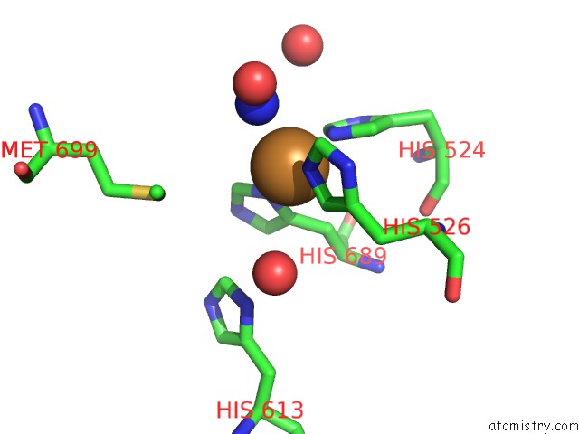
Mono view
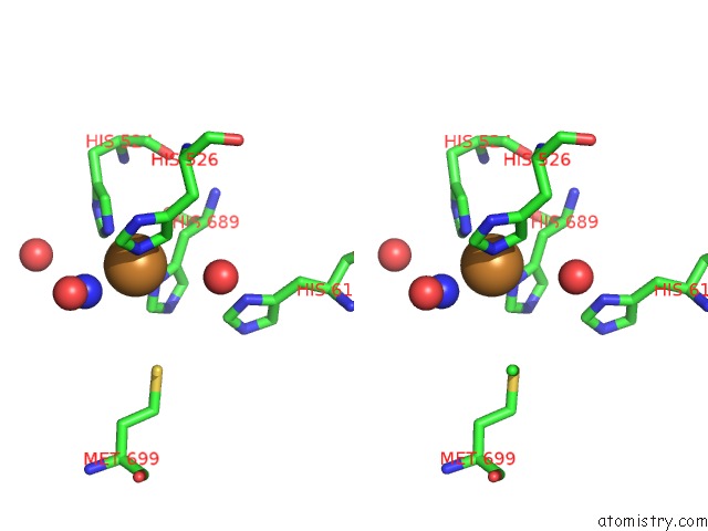
Stereo pair view
Mono view
Stereo pair view
A full contact list of Copper with other atoms in the Cu binding
site number 1 of Crystal Structure of E. Coli Copper-Containing Amine Oxidase Anaerobically Reduced with Beta-Phenylethylamine and Complexed with Nitric Oxide. within 5.0Å range:
|
Copper binding site 2 out of 2 in 1d6y
Go back to
Copper binding site 2 out
of 2 in the Crystal Structure of E. Coli Copper-Containing Amine Oxidase Anaerobically Reduced with Beta-Phenylethylamine and Complexed with Nitric Oxide.
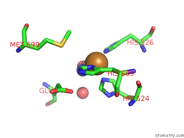
Mono view
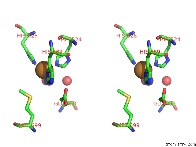
Stereo pair view
Mono view
Stereo pair view
A full contact list of Copper with other atoms in the Cu binding
site number 2 of Crystal Structure of E. Coli Copper-Containing Amine Oxidase Anaerobically Reduced with Beta-Phenylethylamine and Complexed with Nitric Oxide. within 5.0Å range:
|
Reference:
C.M.Wilmot,
J.Hajdu,
M.J.Mcpherson,
P.F.Knowles,
S.E.Phillips.
Visualization of Dioxygen Bound to Copper During Enzyme Catalysis. Science V. 286 1724 1999.
ISSN: ISSN 0036-8075
PubMed: 10576737
DOI: 10.1126/SCIENCE.286.5445.1724
Page generated: Sun Jul 13 23:35:34 2025
ISSN: ISSN 0036-8075
PubMed: 10576737
DOI: 10.1126/SCIENCE.286.5445.1724
Last articles
Fe in 2YXOFe in 2YRS
Fe in 2YXC
Fe in 2YNM
Fe in 2YVJ
Fe in 2YP1
Fe in 2YU2
Fe in 2YU1
Fe in 2YQB
Fe in 2YOO