Copper »
PDB 1bex-1eqw »
1bex »
Copper in PDB 1bex: Structure of Ruthenium-Modified Pseudomonas Aeruginosa Azurin
Protein crystallography data
The structure of Structure of Ruthenium-Modified Pseudomonas Aeruginosa Azurin, PDB code: 1bex
was solved by
S.Faham,
M.W.Day,
D.C.Rees,
with X-Ray Crystallography technique. A brief refinement statistics is given in the table below:
| Resolution Low / High (Å) | 20.00 / 2.30 |
| Space group | C 1 2 1 |
| Cell size a, b, c (Å), α, β, γ (°) | 100.600, 35.400, 74.700, 90.00, 106.50, 90.00 |
| R / Rfree (%) | 20.9 / 28.9 |
Other elements in 1bex:
The structure of Structure of Ruthenium-Modified Pseudomonas Aeruginosa Azurin also contains other interesting chemical elements:
| Ruthenium | (Ru) | 2 atoms |
Copper Binding Sites:
The binding sites of Copper atom in the Structure of Ruthenium-Modified Pseudomonas Aeruginosa Azurin
(pdb code 1bex). This binding sites where shown within
5.0 Angstroms radius around Copper atom.
In total 4 binding sites of Copper where determined in the Structure of Ruthenium-Modified Pseudomonas Aeruginosa Azurin, PDB code: 1bex:
Jump to Copper binding site number: 1; 2; 3; 4;
In total 4 binding sites of Copper where determined in the Structure of Ruthenium-Modified Pseudomonas Aeruginosa Azurin, PDB code: 1bex:
Jump to Copper binding site number: 1; 2; 3; 4;
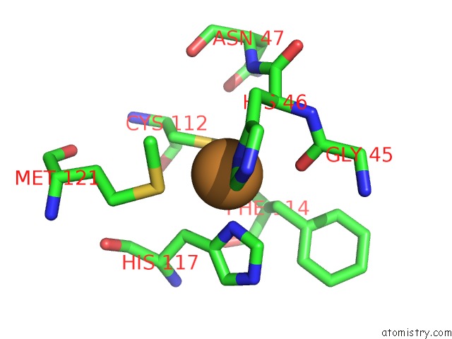
Copper binding site 1 out of 4 in 1bex
Go back to
Copper binding site 1 out
of 4 in the Structure of Ruthenium-Modified Pseudomonas Aeruginosa Azurin
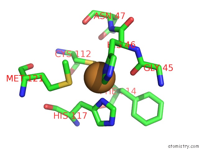
Mono view
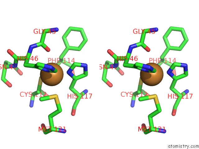
Stereo pair view
Mono view
Stereo pair view
A full contact list of Copper with other atoms in the Cu binding
site number 1 of Structure of Ruthenium-Modified Pseudomonas Aeruginosa Azurin within 5.0Å range:
|
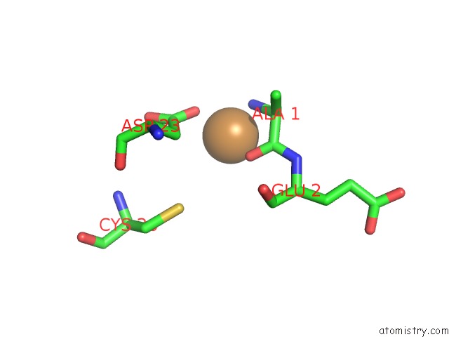
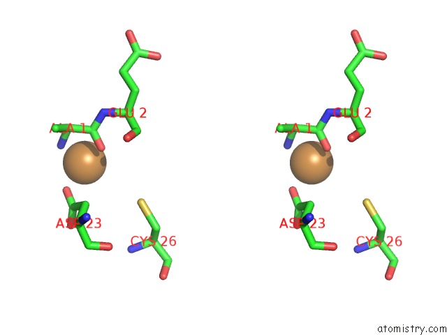
Copper binding site 2 out of 4 in 1bex
Go back to
Copper binding site 2 out
of 4 in the Structure of Ruthenium-Modified Pseudomonas Aeruginosa Azurin
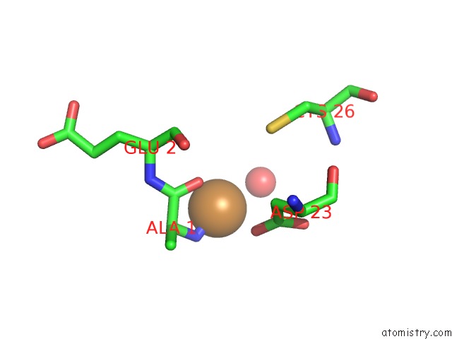
Mono view
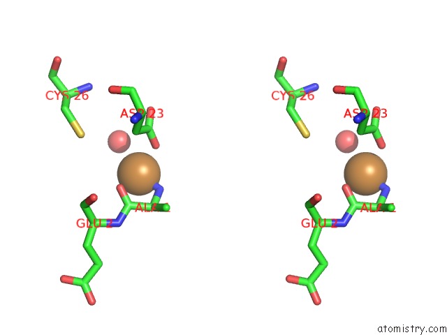
Stereo pair view
Mono view
Stereo pair view
A full contact list of Copper with other atoms in the Cu binding
site number 2 of Structure of Ruthenium-Modified Pseudomonas Aeruginosa Azurin within 5.0Å range:
|
Copper binding site 3 out of 4 in 1bex
Go back to
Copper binding site 3 out
of 4 in the Structure of Ruthenium-Modified Pseudomonas Aeruginosa Azurin
Mono view

Stereo pair view
Mono view
Stereo pair view
A full contact list of Copper with other atoms in the Cu binding
site number 3 of Structure of Ruthenium-Modified Pseudomonas Aeruginosa Azurin within 5.0Å range:
|
Copper binding site 4 out of 4 in 1bex
Go back to
Copper binding site 4 out
of 4 in the Structure of Ruthenium-Modified Pseudomonas Aeruginosa Azurin
Mono view
Stereo pair view
Mono view
Stereo pair view
A full contact list of Copper with other atoms in the Cu binding
site number 4 of Structure of Ruthenium-Modified Pseudomonas Aeruginosa Azurin within 5.0Å range:
|
Reference:
S.Faham,
M.W.Day,
W.B.Connick,
B.R.Crane,
A.J.Di Bilio,
W.P.Schaefer,
D.C.Rees,
H.B.Gray.
Structures of Ruthenium-Modified Pseudomonas Aeruginosa Azurin and [Ru(2,2'-Bipyridine)2(Imidazole)2]SO4 X 10H2O. Acta Crystallogr.,Sect.D V. 55 379 1999.
ISSN: ISSN 0907-4449
PubMed: 10089343
DOI: 10.1107/S0907444998010464
Page generated: Tue Jul 30 21:39:20 2024
ISSN: ISSN 0907-4449
PubMed: 10089343
DOI: 10.1107/S0907444998010464
Last articles
Zn in 9J0NZn in 9J0O
Zn in 9J0P
Zn in 9FJX
Zn in 9EKB
Zn in 9C0F
Zn in 9CAH
Zn in 9CH0
Zn in 9CH3
Zn in 9CH1