Copper »
PDB 1a2v-1baw »
1azr »
Copper in PDB 1azr: Crystal Structure of Pseudomonas Aeruginosa Zinc Azurin Mutant ASP47ASP at 2.4 Angstroms Resolution
Protein crystallography data
The structure of Crystal Structure of Pseudomonas Aeruginosa Zinc Azurin Mutant ASP47ASP at 2.4 Angstroms Resolution, PDB code: 1azr
was solved by
L.Sjolin,
Lc.Tsai,
V.Langer,
T.Pascher,
G.Karlsson,
M.Nordling,
H.Nar,
with X-Ray Crystallography technique. A brief refinement statistics is given in the table below:
| Resolution Low / High (Å) | N/A / 2.40 |
| Space group | P 21 21 21 |
| Cell size a, b, c (Å), α, β, γ (°) | 57.810, 81.530, 112.610, 90.00, 90.00, 90.00 |
| R / Rfree (%) | 17.1 / n/a |
Copper Binding Sites:
The binding sites of Copper atom in the Crystal Structure of Pseudomonas Aeruginosa Zinc Azurin Mutant ASP47ASP at 2.4 Angstroms Resolution
(pdb code 1azr). This binding sites where shown within
5.0 Angstroms radius around Copper atom.
In total 4 binding sites of Copper where determined in the Crystal Structure of Pseudomonas Aeruginosa Zinc Azurin Mutant ASP47ASP at 2.4 Angstroms Resolution, PDB code: 1azr:
Jump to Copper binding site number: 1; 2; 3; 4;
In total 4 binding sites of Copper where determined in the Crystal Structure of Pseudomonas Aeruginosa Zinc Azurin Mutant ASP47ASP at 2.4 Angstroms Resolution, PDB code: 1azr:
Jump to Copper binding site number: 1; 2; 3; 4;
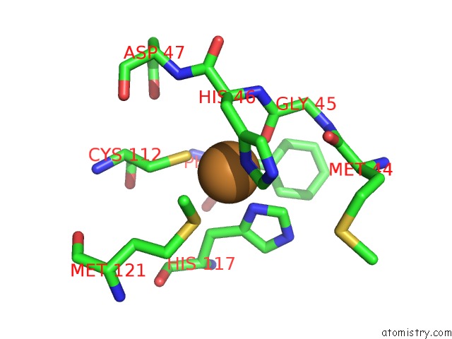
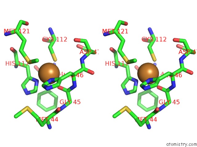
Copper binding site 1 out of 4 in 1azr
Go back to
Copper binding site 1 out
of 4 in the Crystal Structure of Pseudomonas Aeruginosa Zinc Azurin Mutant ASP47ASP at 2.4 Angstroms Resolution
Mono view

Stereo pair view
Mono view
Stereo pair view
A full contact list of Copper with other atoms in the Cu binding
site number 1 of Crystal Structure of Pseudomonas Aeruginosa Zinc Azurin Mutant ASP47ASP at 2.4 Angstroms Resolution within 5.0Å range:
|
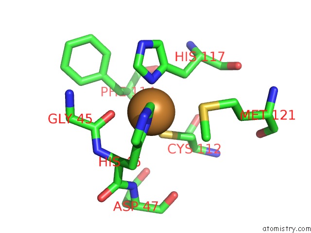
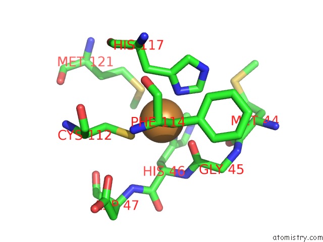
Copper binding site 2 out of 4 in 1azr
Go back to
Copper binding site 2 out
of 4 in the Crystal Structure of Pseudomonas Aeruginosa Zinc Azurin Mutant ASP47ASP at 2.4 Angstroms Resolution
Mono view
Stereo pair view
Mono view
Stereo pair view
A full contact list of Copper with other atoms in the Cu binding
site number 2 of Crystal Structure of Pseudomonas Aeruginosa Zinc Azurin Mutant ASP47ASP at 2.4 Angstroms Resolution within 5.0Å range:
|
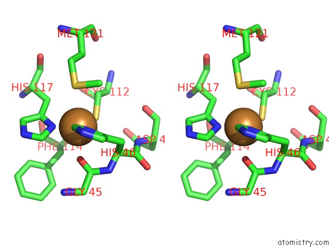
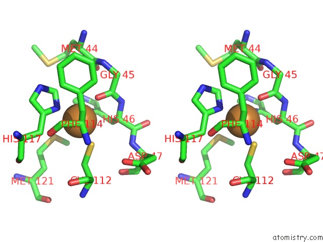
Copper binding site 3 out of 4 in 1azr
Go back to
Copper binding site 3 out
of 4 in the Crystal Structure of Pseudomonas Aeruginosa Zinc Azurin Mutant ASP47ASP at 2.4 Angstroms Resolution
Mono view
Stereo pair view
Mono view
Stereo pair view
A full contact list of Copper with other atoms in the Cu binding
site number 3 of Crystal Structure of Pseudomonas Aeruginosa Zinc Azurin Mutant ASP47ASP at 2.4 Angstroms Resolution within 5.0Å range:
|
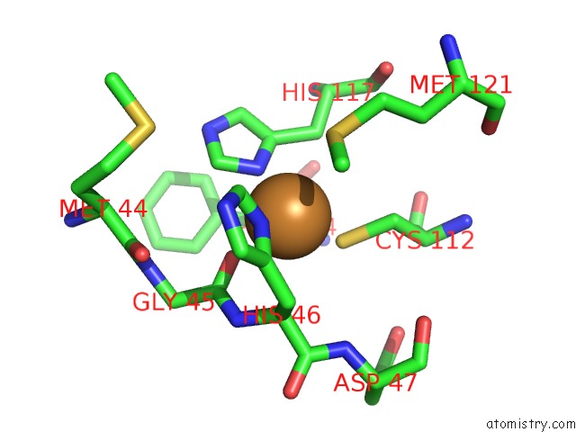
Copper binding site 4 out of 4 in 1azr
Go back to
Copper binding site 4 out
of 4 in the Crystal Structure of Pseudomonas Aeruginosa Zinc Azurin Mutant ASP47ASP at 2.4 Angstroms Resolution
Mono view
Stereo pair view
Mono view
Stereo pair view
A full contact list of Copper with other atoms in the Cu binding
site number 4 of Crystal Structure of Pseudomonas Aeruginosa Zinc Azurin Mutant ASP47ASP at 2.4 Angstroms Resolution within 5.0Å range:
|
Reference:
L.Sjolin,
L.C.Tsai,
V.Langer,
T.Pascher,
G.Karlsson,
M.Nordling,
H.Nar.
Structure of Pseudomonas Aeruginosai Zinc Azurin Mutant ASN47ASP at 2.4 A Resolution. Acta Crystallogr.,Sect.D V. 49 449 1993.
ISSN: ISSN 0907-4449
PubMed: 15299504
DOI: 10.1107/S0907444993005207
Page generated: Tue Jul 30 21:36:24 2024
ISSN: ISSN 0907-4449
PubMed: 15299504
DOI: 10.1107/S0907444993005207
Last articles
Zn in 9MJ5Zn in 9HNW
Zn in 9G0L
Zn in 9FNE
Zn in 9DZN
Zn in 9E0I
Zn in 9D32
Zn in 9DAK
Zn in 8ZXC
Zn in 8ZUF