Copper »
PDB 1a2v-1baw »
1aqp »
Copper in PDB 1aqp: Ribonuclease A Copper Complex
Enzymatic activity of Ribonuclease A Copper Complex
All present enzymatic activity of Ribonuclease A Copper Complex:
3.1.27.5;
3.1.27.5;
Protein crystallography data
The structure of Ribonuclease A Copper Complex, PDB code: 1aqp
was solved by
N.Ramasubbu,
with X-Ray Crystallography technique. A brief refinement statistics is given in the table below:
| Resolution Low / High (Å) | 8.00 / 2.00 |
| Space group | C 1 2 1 |
| Cell size a, b, c (Å), α, β, γ (°) | 58.180, 53.800, 42.900, 90.00, 118.90, 90.00 |
| R / Rfree (%) | 20 / n/a |
Copper Binding Sites:
The binding sites of Copper atom in the Ribonuclease A Copper Complex
(pdb code 1aqp). This binding sites where shown within
5.0 Angstroms radius around Copper atom.
In total 2 binding sites of Copper where determined in the Ribonuclease A Copper Complex, PDB code: 1aqp:
Jump to Copper binding site number: 1; 2;
In total 2 binding sites of Copper where determined in the Ribonuclease A Copper Complex, PDB code: 1aqp:
Jump to Copper binding site number: 1; 2;
Copper binding site 1 out of 2 in 1aqp
Go back to
Copper binding site 1 out
of 2 in the Ribonuclease A Copper Complex
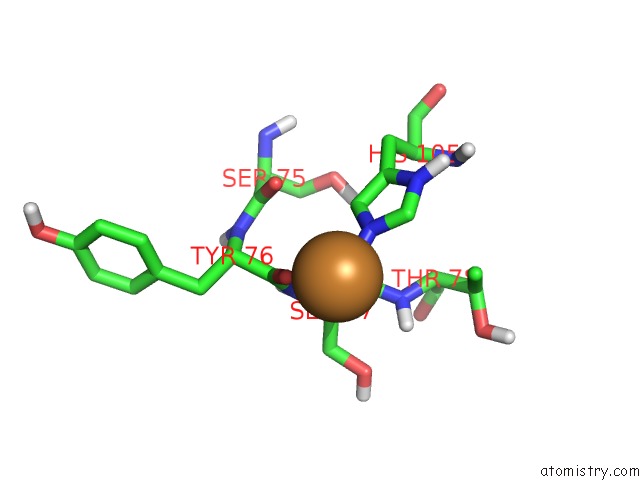
Mono view
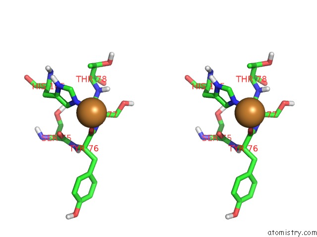
Stereo pair view
Mono view
Stereo pair view
A full contact list of Copper with other atoms in the Cu binding
site number 1 of Ribonuclease A Copper Complex within 5.0Å range:
|
Copper binding site 2 out of 2 in 1aqp
Go back to
Copper binding site 2 out
of 2 in the Ribonuclease A Copper Complex
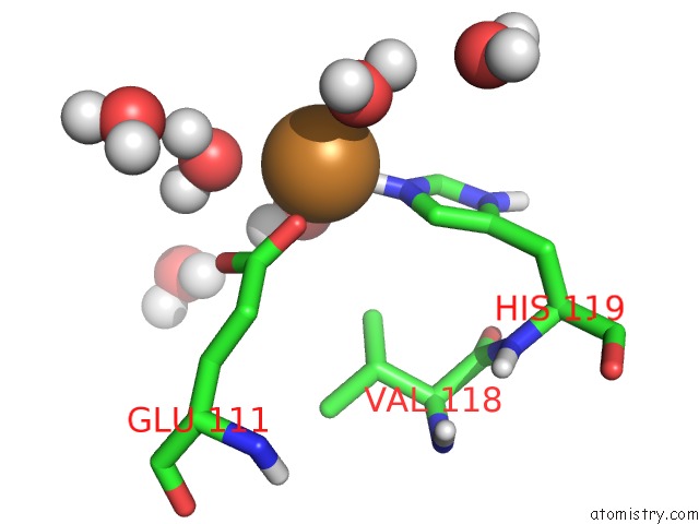
Mono view
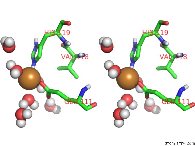
Stereo pair view
Mono view
Stereo pair view
A full contact list of Copper with other atoms in the Cu binding
site number 2 of Ribonuclease A Copper Complex within 5.0Å range:
|
Reference:
R.Balakrishnan,
N.Ramasubbu,
K.I.Varughese,
R.Parthasarathy.
Crystal Structures of the Copper and Nickel Complexes of Rnase A: Metal-Induced Interprotein Interactions and Identification of A Novel Copper Binding Motif. Proc.Natl.Acad.Sci.Usa V. 94 9620 1997.
ISSN: ISSN 0027-8424
PubMed: 9275172
DOI: 10.1073/PNAS.94.18.9620
Page generated: Tue Jul 30 21:29:31 2024
ISSN: ISSN 0027-8424
PubMed: 9275172
DOI: 10.1073/PNAS.94.18.9620
Last articles
Ba in 2GPXBa in 1U9S
Ba in 284D
Ba in 2DQO
Ba in 2BOU
Ba in 2CIS
Ba in 2ADI
Ba in 2B5E
Ba in 1ZQN
Ba in 1Y6S