Copper »
PDB 1a2v-1baw »
1ag6 »
Copper in PDB 1ag6: Plastocyanin From Spinach
Protein crystallography data
The structure of Plastocyanin From Spinach, PDB code: 1ag6
was solved by
Y.Xue,
M.Okvist,
S.Young,
with X-Ray Crystallography technique. A brief refinement statistics is given in the table below:
| Resolution Low / High (Å) | 30.00 / 1.60 |
| Space group | P 31 2 1 |
| Cell size a, b, c (Å), α, β, γ (°) | 75.582, 75.582, 32.707, 90.00, 90.00, 120.00 |
| R / Rfree (%) | 19.1 / 22.4 |
Copper Binding Sites:
The binding sites of Copper atom in the Plastocyanin From Spinach
(pdb code 1ag6). This binding sites where shown within
5.0 Angstroms radius around Copper atom.
In total only one binding site of Copper was determined in the Plastocyanin From Spinach, PDB code: 1ag6:
In total only one binding site of Copper was determined in the Plastocyanin From Spinach, PDB code: 1ag6:
Copper binding site 1 out of 1 in 1ag6
Go back to
Copper binding site 1 out
of 1 in the Plastocyanin From Spinach
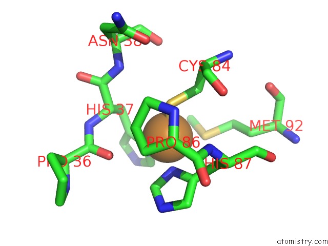
Mono view
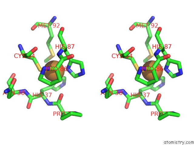
Stereo pair view
Mono view
Stereo pair view
A full contact list of Copper with other atoms in the Cu binding
site number 1 of Plastocyanin From Spinach within 5.0Å range:
|
Reference:
Y.Xue,
M.Okvist,
O.Hansson,
S.Young.
Crystal Structure of Spinach Plastocyanin at 1.7 A Resolution. Protein Sci. V. 7 2099 1998.
ISSN: ISSN 0961-8368
PubMed: 9792096
Page generated: Tue Jul 30 21:28:38 2024
ISSN: ISSN 0961-8368
PubMed: 9792096
Last articles
Cl in 3GDXCl in 3GC8
Cl in 3GD2
Cl in 3GCS
Cl in 3GC7
Cl in 3GB4
Cl in 3GC2
Cl in 3GBA
Cl in 3GBN
Cl in 3GAE