Copper »
PDB 1a2v-1baw »
1aan »
Copper in PDB 1aan: Crystal Structure Analysis of Amicyanin and Apoamicyanin From Paracoccus Denitrificans at 2.0 Angstroms and 1.8 Angstroms Resolution
Protein crystallography data
The structure of Crystal Structure Analysis of Amicyanin and Apoamicyanin From Paracoccus Denitrificans at 2.0 Angstroms and 1.8 Angstroms Resolution, PDB code: 1aan
was solved by
L.Chen,
R.C.E.Durley,
L.W.Lim,
F.S.Mathews,
with X-Ray Crystallography technique. A brief refinement statistics is given in the table below:
| Resolution Low / High (Å) | N/A / 2.00 |
| Space group | P 1 21 1 |
| Cell size a, b, c (Å), α, β, γ (°) | 28.950, 56.540, 27.550, 90.00, 96.38, 90.00 |
| R / Rfree (%) | 15.7 / n/a |
Copper Binding Sites:
The binding sites of Copper atom in the Crystal Structure Analysis of Amicyanin and Apoamicyanin From Paracoccus Denitrificans at 2.0 Angstroms and 1.8 Angstroms Resolution
(pdb code 1aan). This binding sites where shown within
5.0 Angstroms radius around Copper atom.
In total only one binding site of Copper was determined in the Crystal Structure Analysis of Amicyanin and Apoamicyanin From Paracoccus Denitrificans at 2.0 Angstroms and 1.8 Angstroms Resolution, PDB code: 1aan:
In total only one binding site of Copper was determined in the Crystal Structure Analysis of Amicyanin and Apoamicyanin From Paracoccus Denitrificans at 2.0 Angstroms and 1.8 Angstroms Resolution, PDB code: 1aan:
Copper binding site 1 out of 1 in 1aan
Go back to
Copper binding site 1 out
of 1 in the Crystal Structure Analysis of Amicyanin and Apoamicyanin From Paracoccus Denitrificans at 2.0 Angstroms and 1.8 Angstroms Resolution
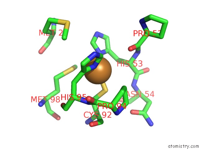
Mono view
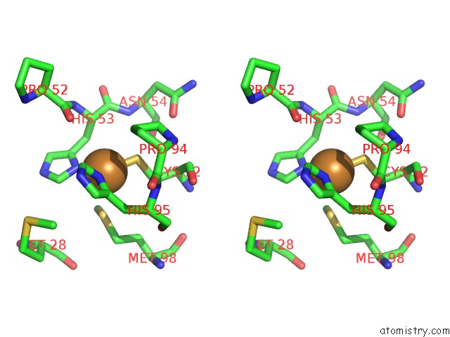
Stereo pair view
Mono view
Stereo pair view
A full contact list of Copper with other atoms in the Cu binding
site number 1 of Crystal Structure Analysis of Amicyanin and Apoamicyanin From Paracoccus Denitrificans at 2.0 Angstroms and 1.8 Angstroms Resolution within 5.0Å range:
|
Reference:
R.Durley,
L.Chen,
L.W.Lim,
F.S.Mathews,
V.L.Davidson.
Crystal Structure Analysis of Amicyanin and Apoamicyanin From Paracoccus Denitrificans at 2.0 A and 1.8 A Resolution. Protein Sci. V. 2 739 1993.
ISSN: ISSN 0961-8368
PubMed: 8495197
Page generated: Sun Jul 13 23:24:33 2025
ISSN: ISSN 0961-8368
PubMed: 8495197
Last articles
F in 4IVMF in 4IV2
F in 4IUI
F in 4IN4
F in 4IU7
F in 4ITI
F in 4IUE
F in 4IRU
F in 4ITJ
F in 4ISF