Copper »
PDB 1a2v-1baw »
1a8z »
Copper in PDB 1a8z: Structure Determination of A 16.8KDA Copper Protein Rusticyanin at 2.1A Resolution Using Anomalous Scattering Data with Direct Methods
Protein crystallography data
The structure of Structure Determination of A 16.8KDA Copper Protein Rusticyanin at 2.1A Resolution Using Anomalous Scattering Data with Direct Methods, PDB code: 1a8z
was solved by
I.Harvey,
Q.Hao,
E.M.H.Duke,
W.J.Ingledew,
S.S.Hasnain,
with X-Ray Crystallography technique. A brief refinement statistics is given in the table below:
| Resolution Low / High (Å) | 8.00 / 2.10 |
| Space group | P 1 21 1 |
| Cell size a, b, c (Å), α, β, γ (°) | 32.430, 60.680, 38.010, 90.00, 107.82, 90.00 |
| R / Rfree (%) | 18.7 / 21.9 |
Copper Binding Sites:
The binding sites of Copper atom in the Structure Determination of A 16.8KDA Copper Protein Rusticyanin at 2.1A Resolution Using Anomalous Scattering Data with Direct Methods
(pdb code 1a8z). This binding sites where shown within
5.0 Angstroms radius around Copper atom.
In total only one binding site of Copper was determined in the Structure Determination of A 16.8KDA Copper Protein Rusticyanin at 2.1A Resolution Using Anomalous Scattering Data with Direct Methods, PDB code: 1a8z:
In total only one binding site of Copper was determined in the Structure Determination of A 16.8KDA Copper Protein Rusticyanin at 2.1A Resolution Using Anomalous Scattering Data with Direct Methods, PDB code: 1a8z:
Copper binding site 1 out of 1 in 1a8z
Go back to
Copper binding site 1 out
of 1 in the Structure Determination of A 16.8KDA Copper Protein Rusticyanin at 2.1A Resolution Using Anomalous Scattering Data with Direct Methods
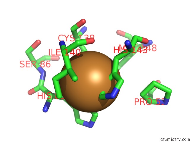
Mono view
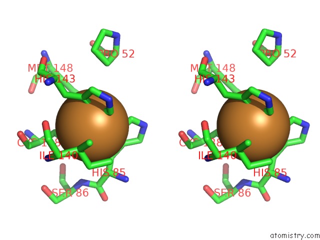
Stereo pair view
Mono view
Stereo pair view
A full contact list of Copper with other atoms in the Cu binding
site number 1 of Structure Determination of A 16.8KDA Copper Protein Rusticyanin at 2.1A Resolution Using Anomalous Scattering Data with Direct Methods within 5.0Å range:
|
Reference:
I.Harvey,
Q.Hao,
E.M.Duke,
W.J.Ingledew,
S.S.Hasnain.
Structure Determination of A 16.8 kDa Copper Protein at 2.1 A Resolution Using Anomalous Scattering Data with Direct Methods. Acta Crystallogr.,Sect.D V. 54 629 1998.
ISSN: ISSN 0907-4449
PubMed: 9761859
DOI: 10.1107/S0907444998005423
Page generated: Tue Jul 30 21:27:41 2024
ISSN: ISSN 0907-4449
PubMed: 9761859
DOI: 10.1107/S0907444998005423
Last articles
Zn in 9MJ5Zn in 9HNW
Zn in 9G0L
Zn in 9FNE
Zn in 9DZN
Zn in 9E0I
Zn in 9D32
Zn in 9DAK
Zn in 8ZXC
Zn in 8ZUF