Copper in PDB 8yk7: Structure of Rib Domain From Surface Adhesin of Limosilactobacillus Reuteri
Protein crystallography data
The structure of Structure of Rib Domain From Surface Adhesin of Limosilactobacillus Reuteri, PDB code: 8yk7
was solved by
Y.Xue,
X.Kang,
with X-Ray Crystallography technique. A brief refinement statistics is given in the table below:
| Resolution Low / High (Å) | 19.91 / 1.35 |
| Space group | P 61 |
| Cell size a, b, c (Å), α, β, γ (°) | 76.274, 76.274, 23.326, 90, 90, 120 |
| R / Rfree (%) | 12.7 / 16.7 |
Other elements in 8yk7:
The structure of Structure of Rib Domain From Surface Adhesin of Limosilactobacillus Reuteri also contains other interesting chemical elements:
| Sodium | (Na) | 1 atom |
Copper Binding Sites:
The binding sites of Copper atom in the Structure of Rib Domain From Surface Adhesin of Limosilactobacillus Reuteri
(pdb code 8yk7). This binding sites where shown within
5.0 Angstroms radius around Copper atom.
In total 2 binding sites of Copper where determined in the Structure of Rib Domain From Surface Adhesin of Limosilactobacillus Reuteri, PDB code: 8yk7:
Jump to Copper binding site number: 1; 2;
In total 2 binding sites of Copper where determined in the Structure of Rib Domain From Surface Adhesin of Limosilactobacillus Reuteri, PDB code: 8yk7:
Jump to Copper binding site number: 1; 2;
Copper binding site 1 out of 2 in 8yk7
Go back to
Copper binding site 1 out
of 2 in the Structure of Rib Domain From Surface Adhesin of Limosilactobacillus Reuteri
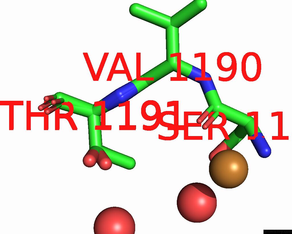
Mono view
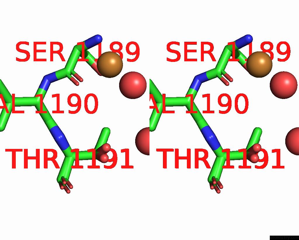
Stereo pair view
Mono view
Stereo pair view
A full contact list of Copper with other atoms in the Cu binding
site number 1 of Structure of Rib Domain From Surface Adhesin of Limosilactobacillus Reuteri within 5.0Å range:
|
Copper binding site 2 out of 2 in 8yk7
Go back to
Copper binding site 2 out
of 2 in the Structure of Rib Domain From Surface Adhesin of Limosilactobacillus Reuteri
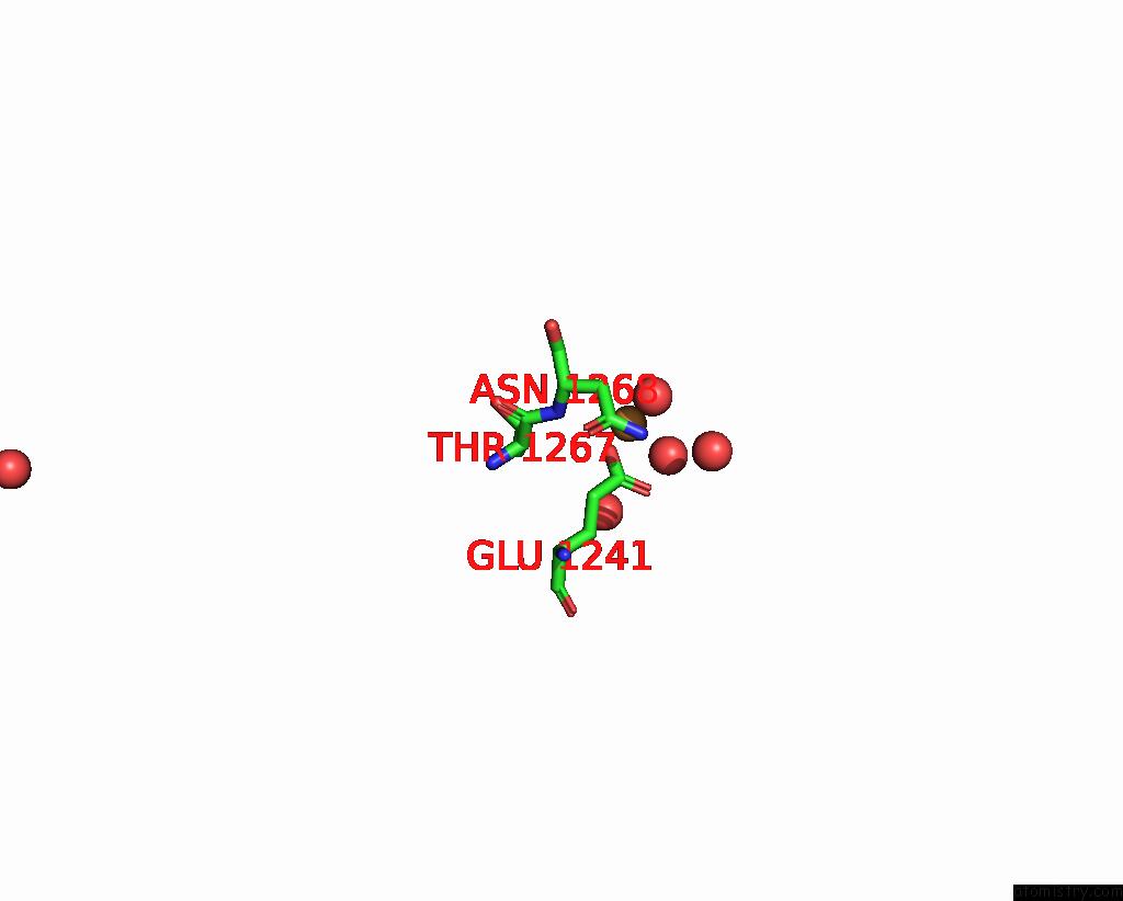
Mono view
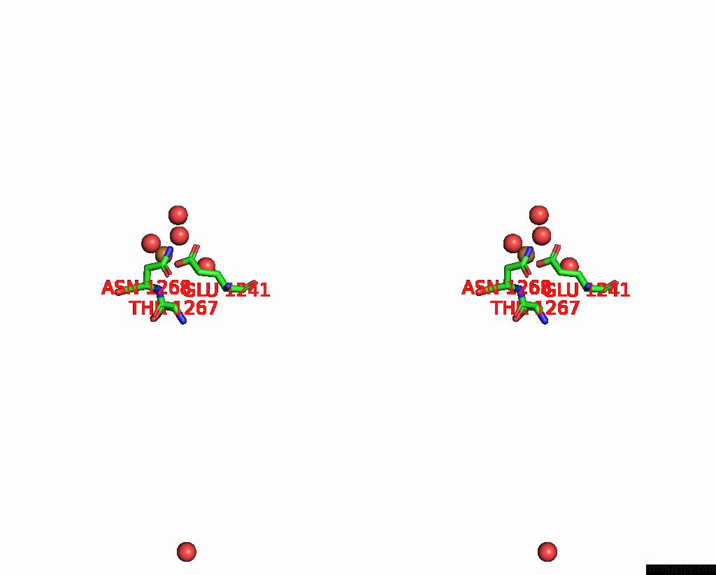
Stereo pair view
Mono view
Stereo pair view
A full contact list of Copper with other atoms in the Cu binding
site number 2 of Structure of Rib Domain From Surface Adhesin of Limosilactobacillus Reuteri within 5.0Å range:
|
Reference:
Y.Xue,
Z.Wu,
X.Kang.
Crystal Structure of the Rib Domain of the Cell-Wall-Anchored Surface Protein From Limosilactobacillus Reuteri. Acta Crystallogr.,Sect.F 2024.
ISSN: ESSN 2053-230X
PubMed: 39196706
DOI: 10.1107/S2053230X24007970
Page generated: Sat Sep 28 19:56:04 2024
ISSN: ESSN 2053-230X
PubMed: 39196706
DOI: 10.1107/S2053230X24007970
Last articles
Zn in 9MJ5Zn in 9HNW
Zn in 9G0L
Zn in 9FNE
Zn in 9DZN
Zn in 9E0I
Zn in 9D32
Zn in 9DAK
Zn in 8ZXC
Zn in 8ZUF