Copper in PDB 6xx3: Crystal Structure of the C-Src SH3 Domain H122R-Q128E Mutant in Complex with Cu(II) at pH 6.5 Co-Crystallized with Methyl Beta- Cyclodextrin
Enzymatic activity of Crystal Structure of the C-Src SH3 Domain H122R-Q128E Mutant in Complex with Cu(II) at pH 6.5 Co-Crystallized with Methyl Beta- Cyclodextrin
All present enzymatic activity of Crystal Structure of the C-Src SH3 Domain H122R-Q128E Mutant in Complex with Cu(II) at pH 6.5 Co-Crystallized with Methyl Beta- Cyclodextrin:
2.7.10.2;
2.7.10.2;
Protein crystallography data
The structure of Crystal Structure of the C-Src SH3 Domain H122R-Q128E Mutant in Complex with Cu(II) at pH 6.5 Co-Crystallized with Methyl Beta- Cyclodextrin, PDB code: 6xx3
was solved by
A.Camara-Artigas,
with X-Ray Crystallography technique. A brief refinement statistics is given in the table below:
| Resolution Low / High (Å) | 17.78 / 1.36 |
| Space group | P 31 2 1 |
| Cell size a, b, c (Å), α, β, γ (°) | 35.566, 35.566, 81.076, 90.00, 90.00, 120.00 |
| R / Rfree (%) | 17.9 / 20.4 |
Copper Binding Sites:
The binding sites of Copper atom in the Crystal Structure of the C-Src SH3 Domain H122R-Q128E Mutant in Complex with Cu(II) at pH 6.5 Co-Crystallized with Methyl Beta- Cyclodextrin
(pdb code 6xx3). This binding sites where shown within
5.0 Angstroms radius around Copper atom.
In total only one binding site of Copper was determined in the Crystal Structure of the C-Src SH3 Domain H122R-Q128E Mutant in Complex with Cu(II) at pH 6.5 Co-Crystallized with Methyl Beta- Cyclodextrin, PDB code: 6xx3:
In total only one binding site of Copper was determined in the Crystal Structure of the C-Src SH3 Domain H122R-Q128E Mutant in Complex with Cu(II) at pH 6.5 Co-Crystallized with Methyl Beta- Cyclodextrin, PDB code: 6xx3:
Copper binding site 1 out of 1 in 6xx3
Go back to
Copper binding site 1 out
of 1 in the Crystal Structure of the C-Src SH3 Domain H122R-Q128E Mutant in Complex with Cu(II) at pH 6.5 Co-Crystallized with Methyl Beta- Cyclodextrin
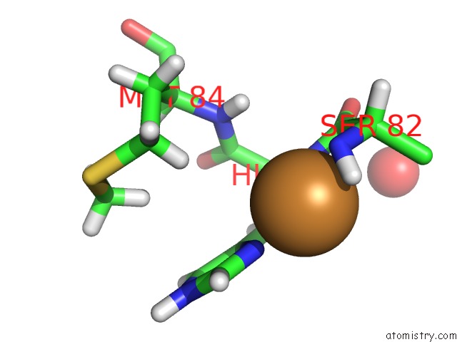
Mono view
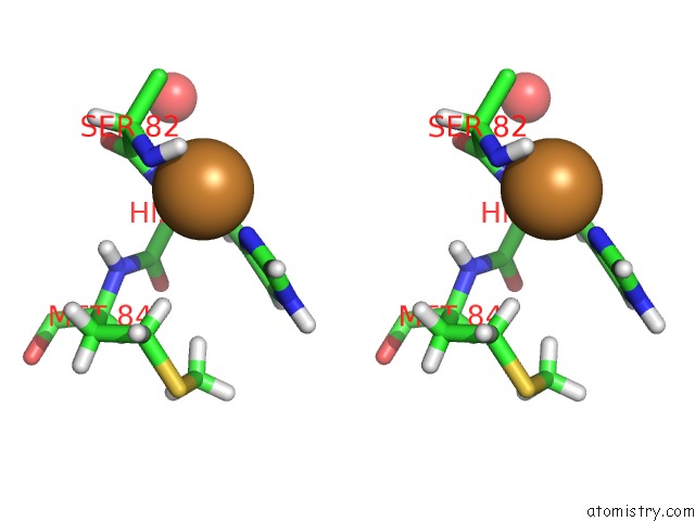
Stereo pair view
Mono view
Stereo pair view
A full contact list of Copper with other atoms in the Cu binding
site number 1 of Crystal Structure of the C-Src SH3 Domain H122R-Q128E Mutant in Complex with Cu(II) at pH 6.5 Co-Crystallized with Methyl Beta- Cyclodextrin within 5.0Å range:
|
Reference:
M.Plaza-Garrido,
M.C.Salinas-Garcia,
J.C.Martinez,
A.Camara-Artigas.
The Effect of An Engineered Atcun Motif on the Structure and Biophysical Properties of the SH3 Domain of C-Src Tyrosine Kinase. J.Biol.Inorg.Chem. 2020.
ISSN: ESSN 1432-1327
PubMed: 32279137
DOI: 10.1007/S00775-020-01785-0
Page generated: Wed Jul 31 07:53:41 2024
ISSN: ESSN 1432-1327
PubMed: 32279137
DOI: 10.1007/S00775-020-01785-0
Last articles
Zn in 9J0NZn in 9J0O
Zn in 9J0P
Zn in 9FJX
Zn in 9EKB
Zn in 9C0F
Zn in 9CAH
Zn in 9CH0
Zn in 9CH3
Zn in 9CH1